


Large Animals
The body size and other anatomic peculiarities of large animals require a highly specialized product range. KARL STORZ has responded with many specifically developed instruments.
- The NEW KARL STORZ Equine Dental Scope
- Snap-in Sheaths from KARL STORZ
- ENDOMAT® SELECT – The Choice is Yours
- Large Animal Videoendoscopes
- Equine Laryngeal Forceps
- Performance Meets Ergonomics: The New DrillCut-X® ARTHRO Shaver Handpiece
- Looking for new endoscopy clients?
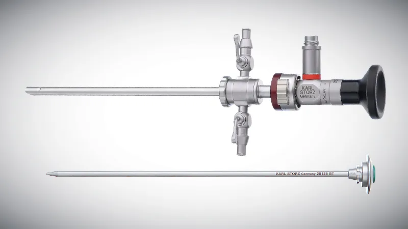
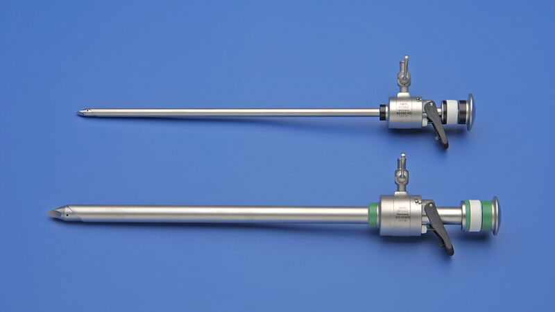
- Extended length TERNAMIAN EndoTIP Cannula for Equine Laparoscopy
- Arthroscopy
- Laparoscopy, Thoracoscopy
- Gastroscopy, Bronchoscopy, Hysteroscopy
- Theloresectoscopy
- Embryo Transfer
- Laparoscopic abomasal repositioning
- VITOM® 25
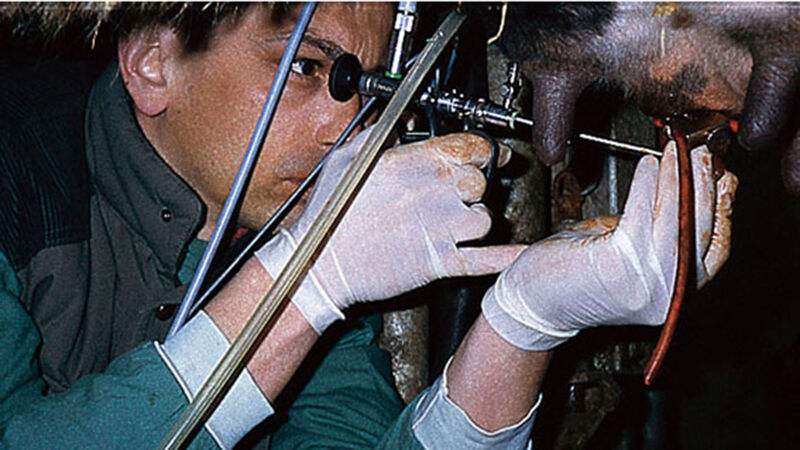
The NEW KARL STORZ Equine Dental Scope

New dimensions are more ergonomic and deliver easier handling
The use of an endoscope during equine dental examinations, cleaning, floating, extractions or other procedures improves visualization and accuracy and allows for documentation of findings.
- Robust construction protects telescope from damage
- Completely soakable
- Light and irrigation connectors are rotatable
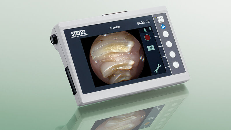
- The lightweight and easy to use C-MAC® HD monitor works with either a standard electrical power cord or with a battery for ease of use in the field or the clinic
- The TELE PACK+ VET is the ultimate mobile endoscopic video system, compatible with all types of endoscopes, including videoendoscopes, rigid telescopes, fiberscopes and exoscopes
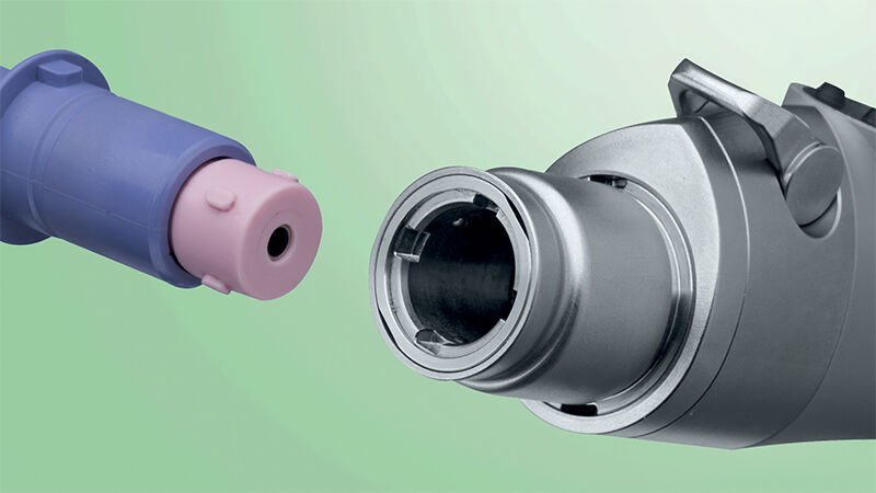
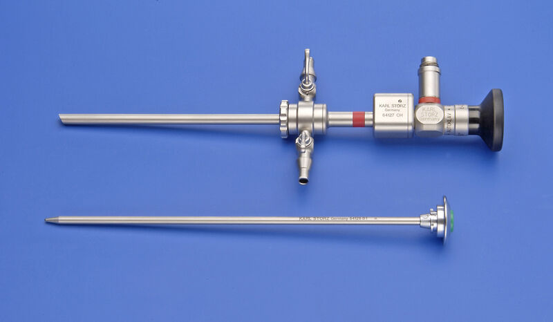
Snap-in Sheaths from KARL STORZ

For a fast, simple and secure telescope-sheath-connection
- The snap-in telescope connection ensures the telescope is much easier to lock into place
- High-flow system for greater flow of irrigation liquid
- Effective cleaning and reprocessing as sheaths can be fully dismantled
ENDOMAT® SELECT – The Choice is Yours
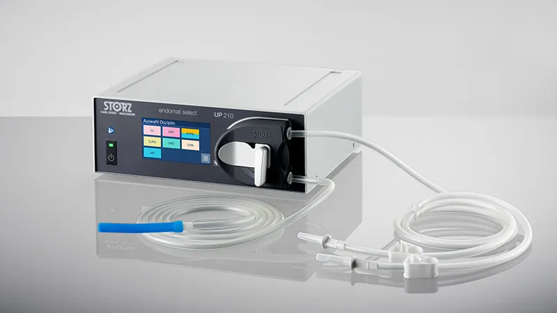
ENDOMAT® SELECT is an interdisciplinary roller pump for the irrigation or suction of fluids during surgical and diagnostic procedures.
Benefits at a glance:
- Cost-efficient pump as modular design supports basic functionality across various fields of application
- Extremely simple handling thanks to tubing set for single hand control
- Safe use thanks to tubing set recognition
- Seamless integration into existing systems
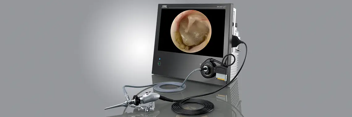
Large Animal Videoendoscopes
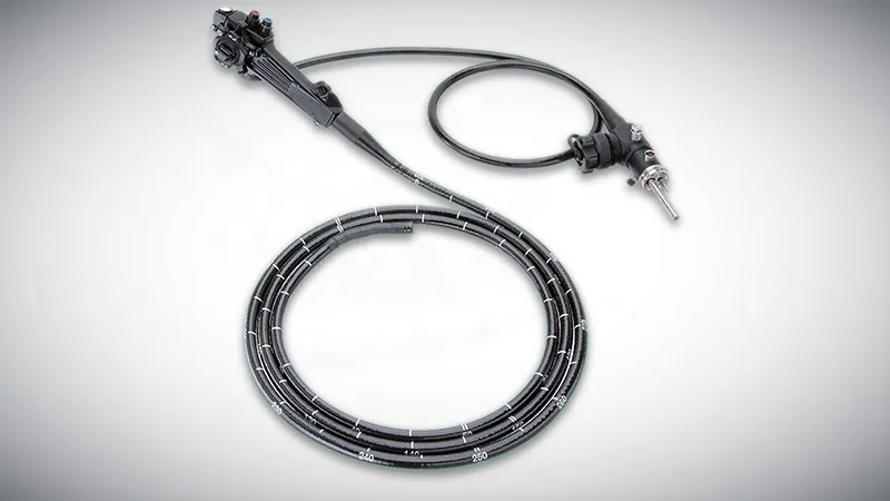
High resolution CCD chip delivers sharp, true color images
- Ideal for numerous large animal applications, including: Gastroscopy, Esophagoscopy, Tracheoscopy, Hysteroscopy, Urethrocystoscopy, Rhinoscopy, Pharyngoscopy and Laryngoscopy
- Large working channel facilitates bigger biopsies to ensure a more accurate diagnosis
- Uniform image brightness, even under difficult lighting conditions
- Large videoendoscopes are compatible with the TELE PACK+ VET camera system
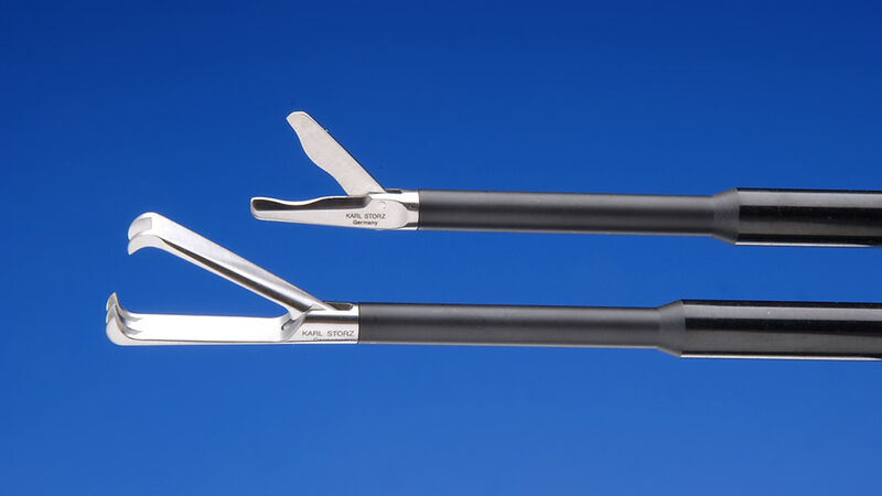
Equine Laryngeal Forceps

Used transnasally alongside KARL STORZ flexible videoendoscopes to perform laryngeal laser surgery
- Curved shaft is ergonomic and allows for smooth passage through the nasal cavity to the larynx
- Ratcheted handle maintains a secure grasp on the larynx for the full duration of the procedure
- Double action jaws
- Large 4 mm jaw width
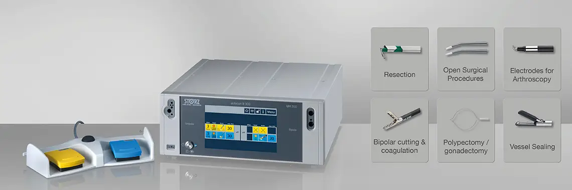
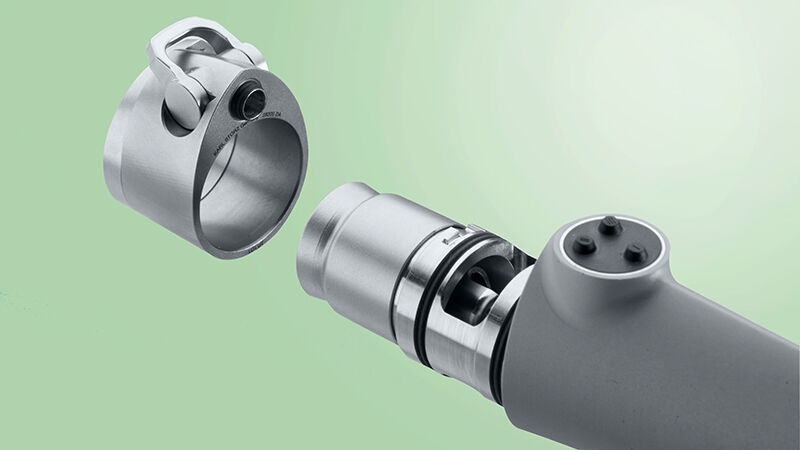
Performance Meets Ergonomics: The New DrillCut-X® ARTHRO Shaver Handpiece
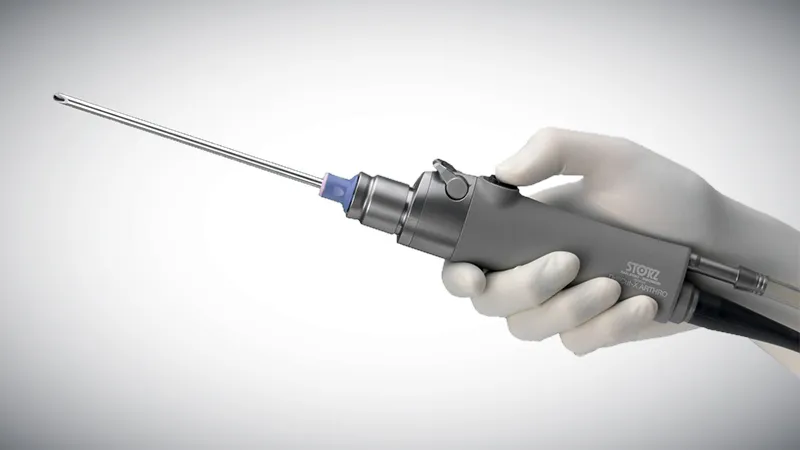
The new DRILLCUT-X® ARTHRO shaver handpiece stands out due to its excellent cleanability and exceptional versatility. Another striking feature is its ergonomic design. Due to its dimensions, the tool is suitable for use in both large and small joints. The shaver handpiece is controlled via the UNIDRIVE® S III ARTHRO console.
- Ergonomic design allows various hand positions
- Possible to lock blades in 4 positions (0°, 90°, 180°, 270°)
- Low-noise operation
- Handpiece weight of only 310 g allows fatigue-free work
Looking for new endoscopy clients?
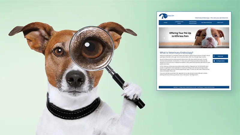
Register at lapspay.com to start receiving FREE referrals for laparoscopy and other endoscopic procedures
- Lapspay.com educates pet owners on the application and benefit of endoscopy in veterinary medicine
- Pet owners can search for vets in their area who offer endoscopy
- Registration is free
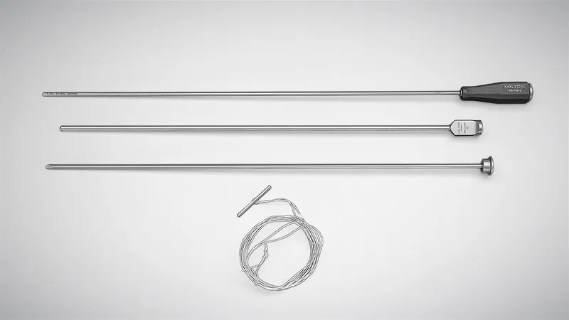
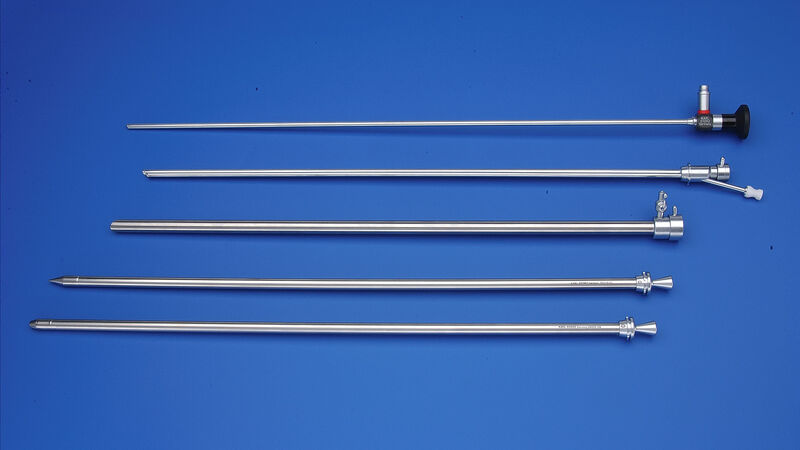
Extended length TERNAMIAN EndoTIP Cannula for Equine Laparoscopy

The longer length makes reaching the abdominal cavity much easier in larger horses
- 20 cm x 11 mm
- Autoclavable
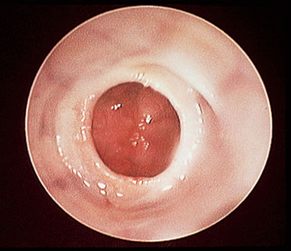
Arthroscopy
Arthroscopy is indispensable in the field of equine orthopedics. With a relatively simple instrument set it is possible to perform diagnostic procedures, which yields information about the location and extent of articular trauma as well as the staging of degenerative joint diseases. Small cartilage defects, synovial hypertrophy, and osteochondrosis can be detected and treated in a single procedure..
Arthroscopy reduces operative time and trauma, enabling the patient to return to normal activity much sooner than with traditional arthrotomy. By distending the joint with fluid or gas, all areas of the articular cartilage can be accessed and debrided under endoscopic visualization.
For further information regarding the instruments and units please see our EndoWorld VET no. 20 and below link "Documentation".
Further Links
Laparoscopy, Thoracoscopy

Laparoscopy has become a desirable alternative to many open abdominal surgical procedures in the horse. Laparoscopic procedures have also been recently developed for cattle, perhaps most significantly for the surgical correction of abomasal displacement.
Depending upon the indication, equine laparoscopy can be performed on patients while standing or in dorsal recumbency. CO2 insufflation is required to distend the abdomen in order to visualize organs. Indications for laparoscopy in the horse include visual examination, organ biopsy, acute or chronic colic, cryptorchidectomy, castration, inguinal hernia, ovariectomy, neoplasia and embryo transfer. KARL STORZ has developed specialized instrumentation for these procedures. The basic set is featured in our ENDOWORLD® VET 22.
Equine thoracoscopy is often performed in the standing horse with pleuropneumonia. In a single procedure, biopsies can be taken and adhesions divided. Thoracoscopy can also be performed in lateral recumbency. Another common indication for thoracoscopy in the horse is thoracic neoplasia. The telescopes and instruments used in thoracoscopy are the same as those used for laparoscopy.
Gastroscopy, Bronchoscopy, Hysteroscopy

In large animal medicine, flexible endoscopes are a standard component of diagnostic equipment. They are particularly useful for differentiating among disease processes in the upper respiratory tract and gastrointestinal tract.
KARL STORZ´s new video endoscope range sets new standards for image quality. Utilization of the very latest in digital processing technology, high resolution CCD chips, and an enhanced optical system results in endoscopes that produce images with unique clarity.
KARL STORZ recommends videoendoscopes with 1.80 m and 2.50 m length for bronchoscopy and 3 m and 3.25 m length for gastroscopy in horses.
The processor of the new Videoendoscope generation is compatible with all rigid and flexibles KARL STORZ optical systems.

The simple, one-handed system consists of a telescope, sheath and resection electrode. Without the aid of an assistant, the clinician can easily maneuver the endoscope so that the operating field is under constant visual control. The KARL STORZ theloresectoscopy set combines the advantages of minimally invasive diagnostics and previously established techniques of electrosurgical resection.

In the standing horse, the instrument pictured below is used to implant the embryo, along with the standard equine laparoscopy instrumentation.
Laparoscopic abomasal repositioning
The increasing number of left abomasal displacements in high-producing cows requires the use of new technologies. Endoscopic techniques reduce treatment times, are more gentle on patients, and hence reduce losses for owners. Laparoscopy additionally facilitates correct toggle placement and combines the advantages of laparotomy (repositioning and fixation under view) with those of the roll-and-suture technique and percutaneous fixation (quick and minimally invasive).
With the animal in a standing position, the laparoscope is placed in the left flank. This positioning ensures a clear view of the displaced abomasum. Under endoscopic guidance, the trocar for toggle placement is inserted into the abomasum. The toggle is fixed in place in the abomasum through the trocar. The procedure can be finished in dorsal recumbency with caudolateral trocar placement, or the instrument for toggle placement can be used with the animal in a standing position. The laparoscope is placed caudolaterally to the sternum, and through a second trocar located caudally to the first, the toggle suture is grasped using a grasping forceps and then tied there. The suture can be cut after 3-4 weeks.
The KARL STORZ abomasopexy set combines an excellent optical system with high quality instruments and toggle. The toggle features 2 suture holes with special rounding to prevent premature suture rupture in the abomasum.
VITOM® 25

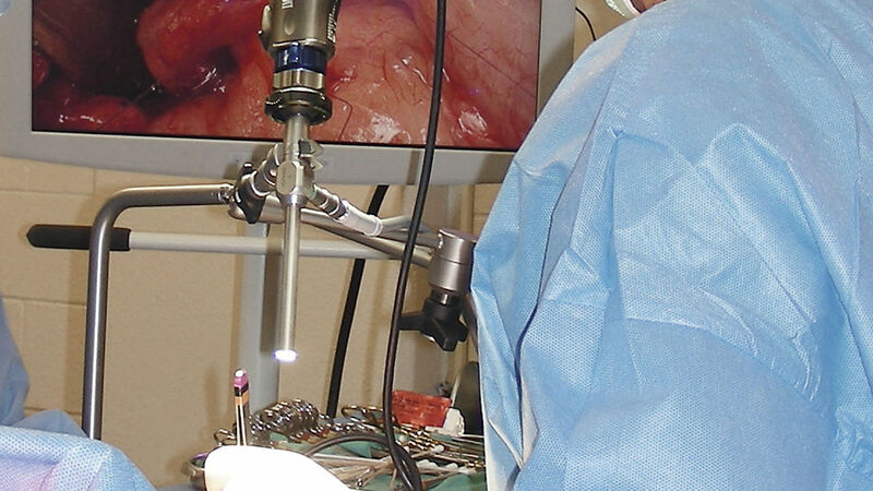
Exoscope for open surgery
The VITOM® 25 exoscope offers a revolutionary new way of displaying open surgical procedures in an ergonomic and high quality manner. Unlike a traditional endoscope, the VITOM® 25 is an “exoscope” which is placed at a distance of 25 to 75 cm from the surgical site, held securely in place by a holding device, giving the surgeon ample workspace.
The magnified image produced by the VITOM® 25 is viewed on a video monitor. This enables the surgeon and support staff to work together comfortably, significantly reducing surgeon fatigue, while the magnification of structures improves surgical precision and accuracy of diagnosis.
This system is ideal for teaching, as is allows for easy, magnified viewing of the surgery, which can also be transmitted to remote locations, without disruption of the procedure.
The ability to capture and archive images and videos of surgical procedures ensures that a detailed and accurate record of the diagnosis and treatment performed is recorded, while creating a valuable tool for training, education, re-examination at a later time, and sharing with clients and colleagues.
The VITOM® 25 works with any existing KARL STORZ video system.