
Sistemas de imagem
A KARL STORZ é pioneira na produção de sistemas de imagens, disponibilizando soluções modulares inovadoras e escaláveis para a obtenção de imagens de excelente qualidade de acordo com as preferências e o orçamento de cada cliente.
A KARL STORZ é pioneira na produção de sistemas de imagens, disponibilizando soluções modulares inovadoras e escaláveis para a obtenção de imagens de excelente qualidade de acordo com as preferências e o orçamento de cada cliente.
BENEFÍCIOS
Precisão. Modularidade. Escalabilidade.
.svg)


Produtos em Destaque


Soluções versáteis de imagiologia
Disponibilizamos sistemas de imagens de excelente qualidade totalmente customizáveis de acordo com as suas preferências e o seu orçamento para utilização em centros cirúrgicos ou em consultórios.
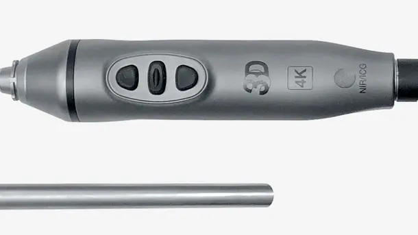
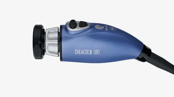
IMAGE1 S™ Rubina®
Alia a tecnologia 3D e 4K com a reprodução de imagens por fluorescência NIR/ICG para facilitar o acesso a informações de excelente qualidade, a identificação de estruturas anatômicas e garantir a precisão dos procedimentos. NIR/ICG amplia as possibilidades de diagnóstico, como a avaliação da perfusão e a detecção do linfonodo sentinela, por exemplo.
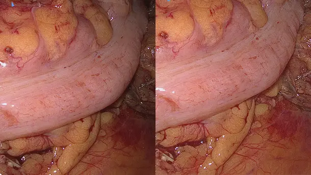
Modos de visualização
Os diferentes modos de visualização facilitam a identificação precisa de estruturas anatômicas.
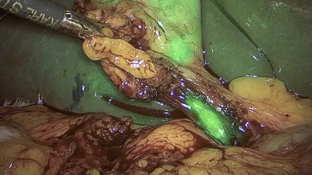
Overlay
Modo que combina a imagem em luz branca padrão com as informações NIR/ICG gerando uma imagem sobreposta verde ou azul.
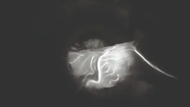
Monocromático
Visualização do sinal NIR/ICG na cor branca em um fundo preto para otimizar a diferenciação de estruturas.
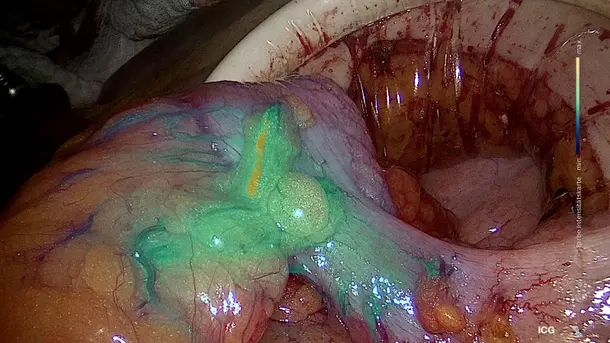
Intensity Map
Visualização da intensidade do sinal NIR/ICG através de uma escala de cores em uma imagem sobreposta.
CLARA
Tecnologia-S que proporciona uma iluminação homogênea, inibe a superexposição e reflexão da imagem e garante melhor visibilidade em áreas escuras.
CHROMA
Tecnologia-S que intensifica o contraste da imagem sem interferir na percepção natural das cores. Isso contribui para tornar as variações de cores e as estruturas anatômicas muito mais nítidas.
CLARA + CHROMA
A combinação entre essas duas Tecnologias-S permite a iluminação homogênea do sítio cirúrgico e realça as estruturas teciduais.
SPECTRA A
Tecnologia-S que facilita a identificação de tecidos através do equilíbrio cromático, utilizando tons verdes e azuis para representar estruturas avermelhadas delicadas, como vasos sanguíneos e mucosas, por exemplo.
SPECTRA B
Equilibra as cores da imagem para reduzir os tons vermelhos e acentuar os tons verdes e azuis. Dessa forma, o fundo da imagem passa a ser exibido na cor esverdeada, evidenciando vasos sanguíneos e capilares.
Portfólio de produtos IMAGE1 S™ Rubina®
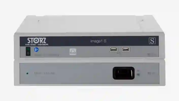
IMAGE 1 S™
Disponibilizamos a solução que você precisa para a visualização ideal do paciente de modo a obter o melhor resultado possível. Nosso portfólio de soluções modulares, sustentáveis e customizadas ampliam as suas opções de visualização.
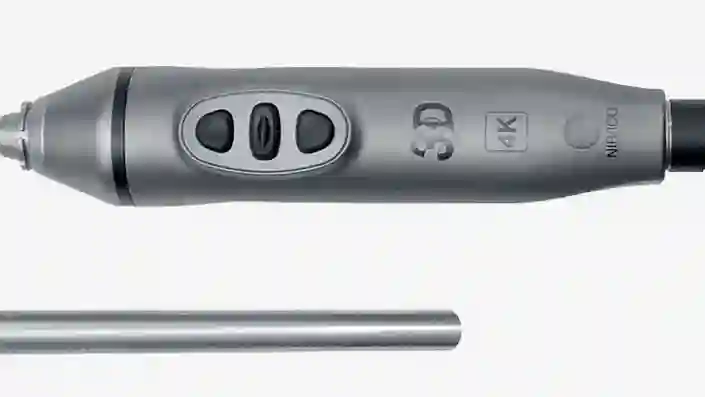
TIPCAM®1 Rubina®
Videoendoscópio "all in one" que alia as tecnologias de última geração 4K, 3D e NIR/ICG em um único produto, proporcionando imagens de excelente qualidade e perfeita percepção de profundidade.
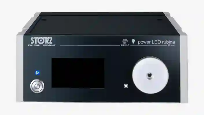
Power LED Rubina®
Fonte de luz fria sem laser muito eficiente e resistente para a utilização em procedimentos com NIR/ICG e no modo de luz branca.
Sistema de imagem NIR/ICG
Configure o sistema utilizando os componentes necessários para a sua prática diária
Áreas de aplicação do IMAGE1 S™ Rubina®
O sistema IMAGE1 S™ Rubina® representa um grande avanço para a realização de cirurgias guiadas por imagens com fluorescência. Deixamos de tatear no "escuro", tendo agora a tecnologia 4K/3D à nossa disposição para oferecer ao nosso paciente o melhor tratamento possível.

IMAGE1 S™ 4U
A tecnologia 4K eleva a qualidade da imagem endoscópica a um outro patamar e é uma importante aliada em nossa busca constante por imagens de excelente qualidade com riqueza de detalhes.
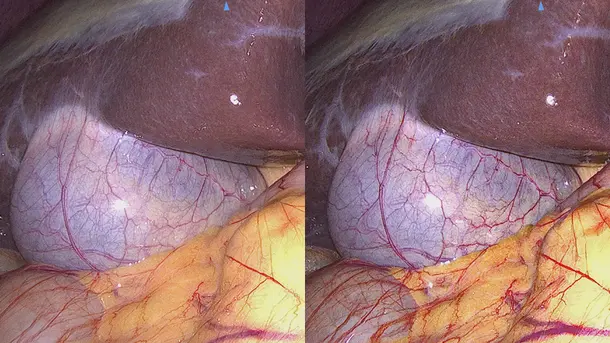
Modos de visualização
Disponibilizamos diferentes modos de visualização para a identificação precisa de estruturas anatômicas.
CLARA
Tecnologia-S que proporciona uma iluminação homogênea, inibe a superexposição e reflexão da imagem e garante melhor visibilidade em áreas escuras.
CHROMA
Tecnologia-S que intensifica o contraste da imagem sem interferir na percepção natural das cores. Isso contribui para tornar as variações de cores e as estruturas anatômicas muito mais nítidas.
CLARA + CHROMA
A combinação entre essas duas Tecnologias-S permite a iluminação homogênea do sítio cirúrgico e realça as estruturas teciduais.
SPECTRA A
Tecnologia-S que facilita a identificação de tecidos através do equilíbrio cromático, utilizando tons verdes e azuis para representar estruturas avermelhadas delicadas, como vasos sanguíneos e mucosas, por exemplo.
SPECTRA B
Equilibra as cores da imagem para reduzir os tons vermelhos e acentuar os tons verdes e azuis. Dessa forma, o fundo da imagem passa a ser exibido na cor esverdeada, evidenciando vasos sanguíneos e capilares.
Portfólio de produtos IMAGE1 S™ 4U
IMAGE 1 S™
Disponibilizamos a solução que você precisa para a visualização ideal do paciente de modo a obter o melhor resultado possível. Nosso portfólio de soluções modulares, sustentáveis e customizadas ampliam as suas opções de visualização.
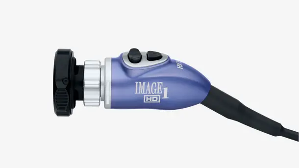
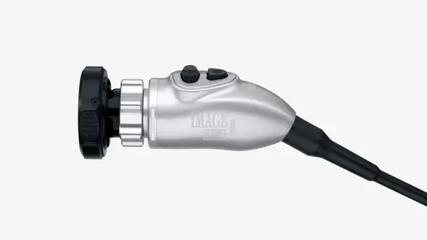
Cabeçote de câmera IMAGE1 S™ 4U
Este cabeçote de câmera é uma ferramenta indispensável para a visualização. A tecnologia 4K é capaz de gerar imagens de alta resolução e ampliar o espaço de cores, facilitando a identificação e a diferenciação de estruturas teciduais.
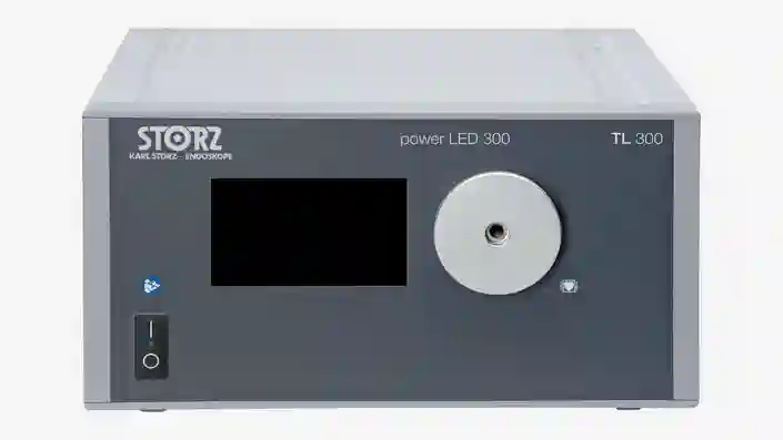
POWER LED 300
Fonte de luz fria sem laser eficiente e resistente indicada para procedimentos no modo de luz branca.
Sistema de imagem 4K
Configure o sistema utilizando os componentes necessários para a sua prática diária.
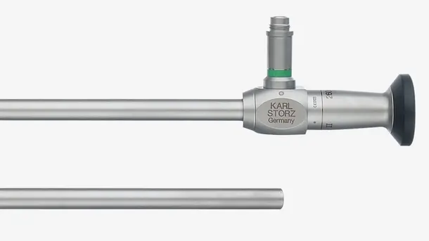
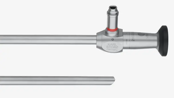
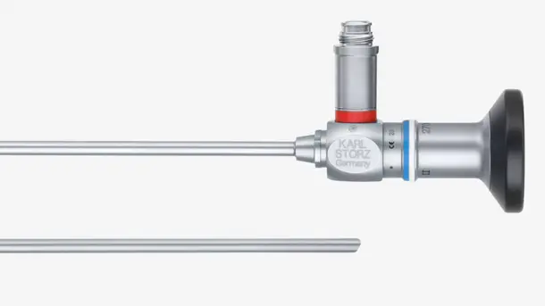
Ópticas HOPKINS®
Nosso portfólio é composto por ópticas compatíveis com nossos sistemas, indicadas para procedimentos com luz branca, para que você disponha de todos os equipamentos necessários para a visualização de imagens de qualidade.
Áreas de aplicação do IMAGE1 S™ 4U
A implementação da tecnologia IMAGE1 S™ em nosso hospital nos permite ver detalhes antes inexplorados. O acesso a imagens de qualidade é de suma importância para o sucesso do tratamento.

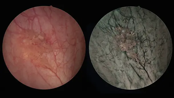
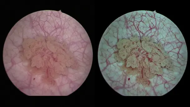
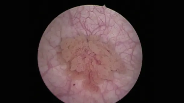
IMAGE1 S™ Saphira™
A técnica Blue Light Imaging (BLI), conhecida até agora como PDD, permite a visualização de tumores malignos em estágio inicial com a aplicação do agente Hexvix/Cysview, normalmente não detectados ou dificilmente detectados com o diagnóstico com luz branca.
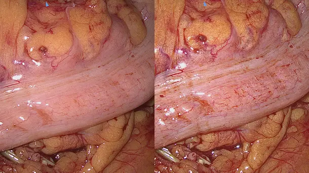
Modos de visualização
Os diferentes modos de visualização facilitam a identificação precisa de estruturas anatômicas.
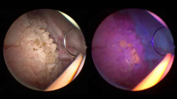
BLI
A luz azul facilita a clara diferenciação entre as células tumorais destacadas em vermelho fluorescente e os tecidos adjacentes visualizados em azul.
CHROMA
Tecnologia-S que intensifica o contraste da imagem, sem interferir na percepção natural das cores. Isso contribui para uma visualização muito mais nítida das tonalidades da imagem e de estruturas anatômicas.
SPECTRA A
Tecnologia-S que facilita a identificação de tecidos através do equilíbrio cromático, utilizando tons verdes e azuis para representar estruturas avermelhadas delicadas, como vasos sanguíneos e mucosas, por exemplo.
SPECTRA B
Equilibra as cores da imagem para reduzir os tons vermelhos e acentuar os tons verdes e azuis. Dessa forma, o fundo da imagem passa a ser exibido na cor esverdeada, evidenciando vasos sanguíneos e capilares.
Portfólio de produtos IMAGE1 S™ Saphira™
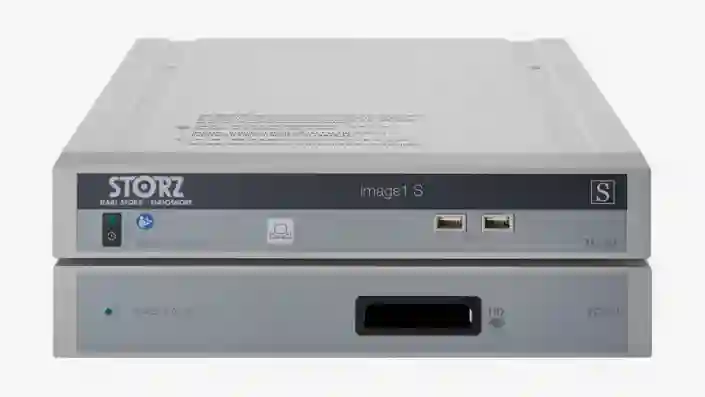
IMAGE 1 S™
Disponibilizamos a solução que você precisa para a visualização ideal do paciente de modo a obter o melhor resultado possível. Nosso portfólio de soluções modulares, sustentáveis e customizadas ampliam as suas opções de visualização.
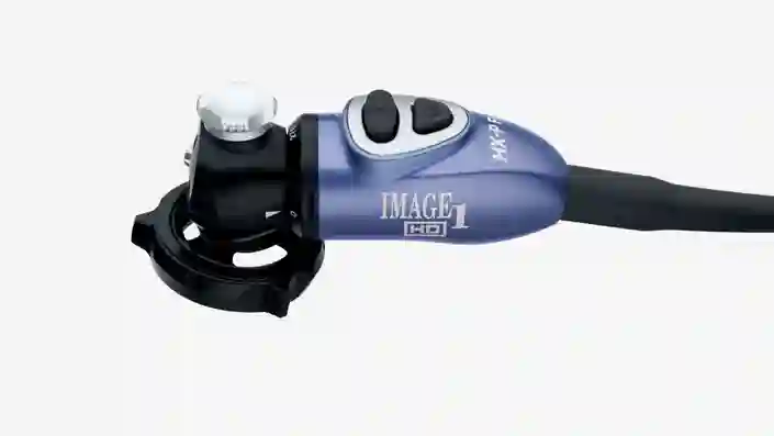
Cabeçote de câmera IMAGE1 S™ HX-P FI
O cabeçote de câmera pendular foi projetado especificamente para aplicações Blue Light Imaging e com luz branca, facilitando o trabalho do especialista por ser um modelo leve e ergonômico.

POWER LED Saphira™
Fonte de luz fria sem laser eficiente e resistente indicada para procedimentos no modo de luz branca e Blue Light Imaging.
Solução Blue Light Imaging
Configure o sistema utilizando os componentes necessários para a sua prática diária.
Áreas de aplicação do IMAGE1 S™ Saphira™
Tive a oportunidade de trabalhar com a nova fonte de luz Saphira™ nos últimos meses e posso afirmar que contribui de maneira significativa para a eficiência dos procedimentos.

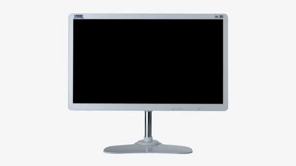
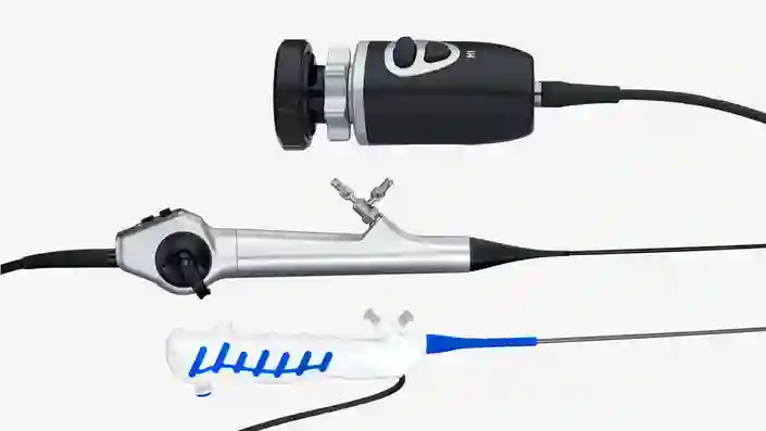
TELE PACK+
Essa plataforma compacta para o diagnóstico e pequenas intervenções pode ser configurada de maneira flexível o que favorece sua utilização em consultórios, clínicas-dia, unidades de urgência e emergência, unidades de tratamento intensivo e no atendimento ambulatorial.
Portfólio de produtos TELE PACK+
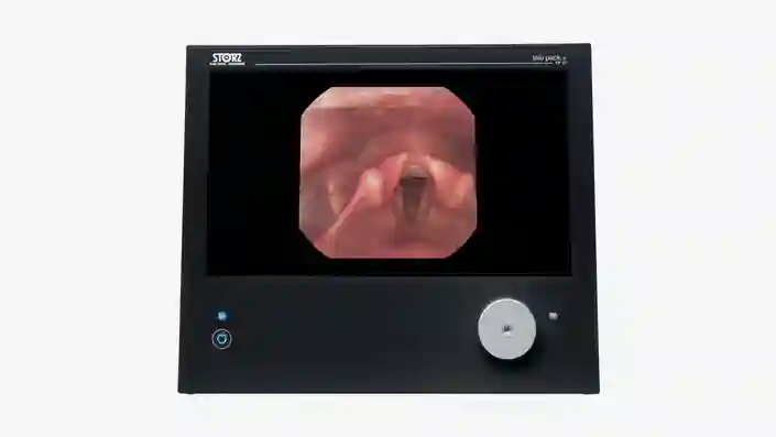
TELE PACK+
Desfrute das vantagens do sistema TELE PACK+ que reúne monitor, fonte de luz LED, unidade de controle de câmera FULL HD e documentação com função de rede integrada em um aparelho portátil e compacto.

X-Line e C-Line
O TELE PACK+ com suas conexões X-Line e C-Line é compatível com uma variedade de endoscópios rígidos, flexíveis e descartáveis, podendo ser utilizado praticamente em quase todas as especialidades médicas.
Disponha de um sistema de imagem FULL HD compacto completo
Configure o sistema utilizando os componentes necessários para a sua prática diária.
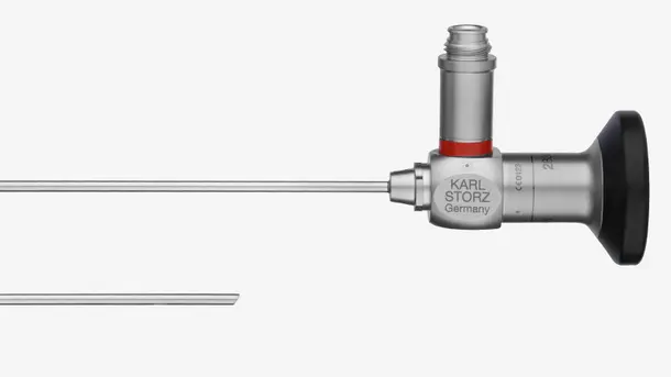
Endoscópios rígidos
Conheça nosso portfólio de ópticas rígidas compatíveis ideais para procedimentos no modo de luz branca.
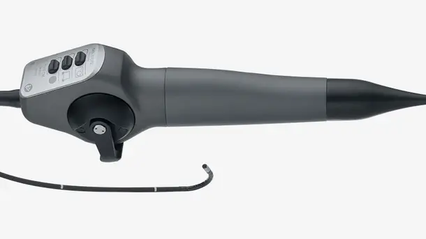
Videoendoscópios
Oferecemos diversos tipos de videoendoscópios flexíveis que permitem a visualização de cavidades estreitas e de difícil acesso, dispensando intervenções invasivas. Nosso vasto portfólio de videoendoscópios compatíveis é indicado para praticamente quase todas as especialidades médicas.
Áreas de aplicação do portfólio TELE PACK+
Preferimos trabalhar com o TELE PACK+ por conta de sua excelente qualidade de imagem que facilita o diagnóstico preciso, mesmo em casos considerados complexos.

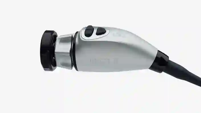
TELECAM C3
A unidade de controle de câmera TELECAM C3 é indicada para a realização de diagnósticos e pequenas intervenções em consultórios médicos e em ambiente hospitalar. Pode ser utilizada em praticamente quase todas as especialidades médicas por ser compatível com diferentes tipos de endoscópios.
Portfólio de produtos TELECAM C3
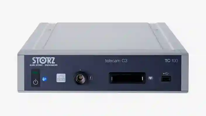
TELECAM C3
Nossa unidade de controle de câmera compacta e econômica foi projetada para a utilização em intervenções endoscópicas simples. Por dispor de duas entradas para câmera permitem a utilização do equipamento com vários tipos de endoscópios.
X-Line e C-Line
As conexões X-Line e C-Line tornam o TELECAM C3 compatível com uma variedade de endoscópios rígidos, flexíveis e descartáveis, podendo ser utilizado praticamente em quase todas as especialidades médicas.
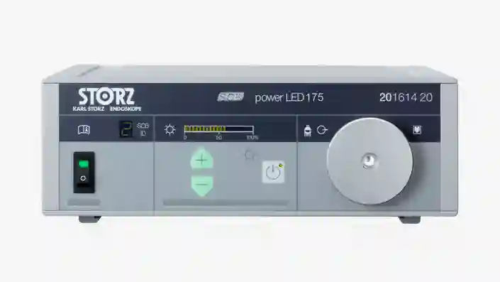
POWER LED 175
Fonte de luz fria sem laser eficiente e resistente que facilita a visualização de cavidades estreitas em procedimentos no modo de luz branca.
Sistema de imagem FULL HD
Configure o sistema utilizando os componentes necessários para a sua prática diária.
Endoscópios rígidos
Conheça nosso portfólio de ópticas rígidas compatíveis ideais para procedimentos no modo de luz branca.
Videoendoscópios
Oferecemos diversos tipos de videoendoscópios flexíveis que permitem a visualização de cavidades estreitas e de difícil acesso, dispensando intervenções invasivas. Nosso vasto portfólio de videoendoscópios compatíveis é indicado para praticamente quase todas as especialidades médicas.
Áreas de aplicação do TELECAM C3
Crie o estado da arte em ambientes cirúrgicos sob medida para suas necessidades
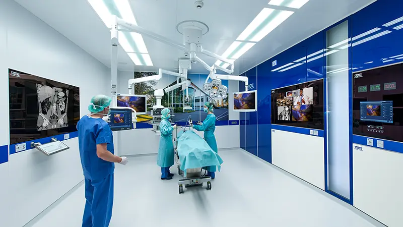
O futuro da sala de cirurgia integrada
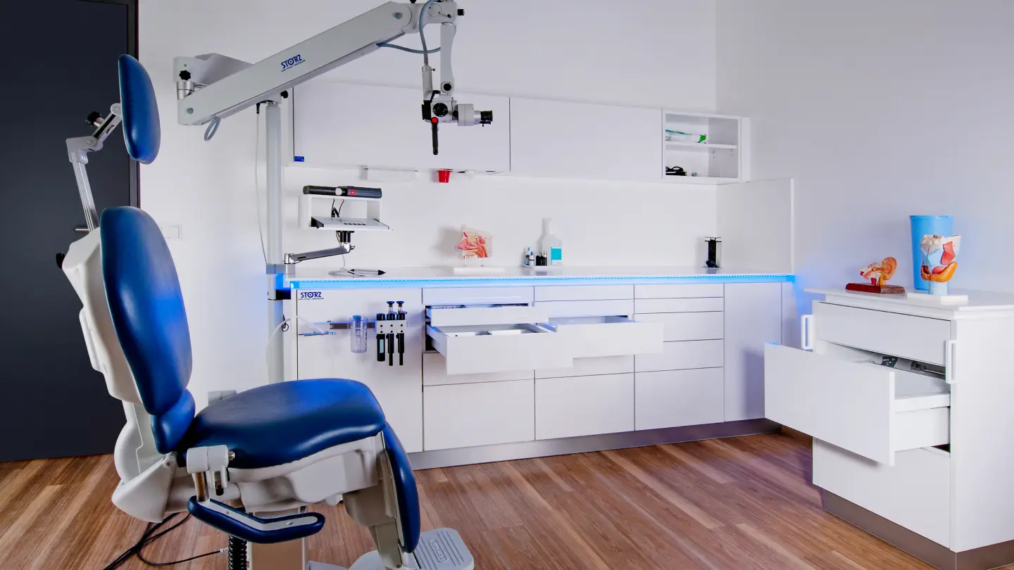
Composição modular do seu consultório
Empoderamento por meio do conhecimento
Entendemos que com o aumento da complexidade das técnicas cirúrgicas, dos instrumentos e dos dispositivos utilizados durante os procedimentos, aumenta também, a importância da formação, especialização e desenvolvimento profissional. Por meio de cursos de capacitação sob medida, workshops e congressos, nos empenhamos para apoiar médicos e assistentes do mundo inteiro.


CONTACT Get in touch with our specialists
We'll guide you through the whole process and help customize the right solution for your surgical needs.