
Imaging
KARL STORZ è un’azienda pioniere nel campo dell’imaging endoscopico che ti offre soluzioni innovative, modulari e scalabili per supportarti con la migliore catena di immagini e soddisfa al meglio le tue esigenze e il tuo budget.
KARL STORZ è un’azienda pioniere nel campo dell’imaging endoscopico che ti offre soluzioni innovative, modulari e scalabili per supportarti con la migliore catena di immagini e soddisfa al meglio le tue esigenze e il tuo budget.
VANTAGGI
Preciso. Modulare. Scalabile.
.svg)


Prodotti in evidenza


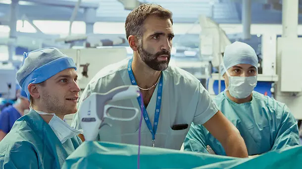
Soluzioni versatili di imaging
Le nostre tecnologie di imaging consentono di ottenere una visualizzazione ottica perfezionata usata sia in sala operatoria che negli studi medici. Con le nostre numerose e opzioni di configurazione puoi combinare in modo individuale la catena di immagini per rispondere al meglio alle tue esigenze e al tuo budget.
IMAGE1 S™ Rubina®
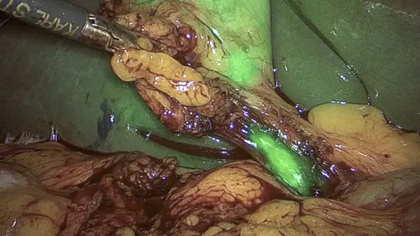
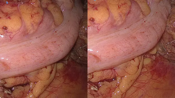
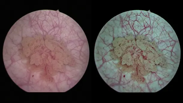
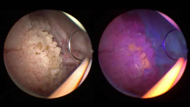
La combinazione delle tecnologie 3D e 4K con l’imaging a fluorescenza NIR/ICG garantisce informazioni di elevata qualità e permette di migliorare la detezione e la precisione chirurgica. NIR/ICG consente di ampliare le possibilità di diagnosi, ad es. la valutazione della perfusione e il riconoscimento dei linfonodi sentinella.
Modalità di visualizzazione
Scopri diverse modalità di visualizzazione per una migliore differenziazione e identificazione delle strutture.
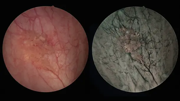
Overlay
In Overlay l’immagine standard a luce bianca è combinata con le informazioni NIR/ICG, generando un’immagine sovrapposta verde o blu.
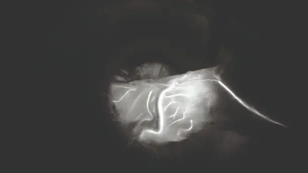
Monochromatic
Il segnale NIR/ICG viene visualizzato in bianco su sfondo nero per ottenere la miglior differenziazione possibile rispetto ai tessuti circostanti.
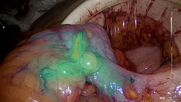
Intensity Map
Consente di visualizzare l’intensità del segnale NIR/ICG utilizzando una scala colorimetrica nell’immagine sovrapposta.
CLARA
Le S-Technology assicurano un’illuminazione omogenea, evitando sovraesposizioni e riflessi e, nello stesso tempo, permettono uno schiarimento delle aeree più in ombra.
CHROMA
Con questa modalità di visualizzazione il contrasto cromatico è esaltato senza alterare la naturale percezione del colore. Le differenze di colore e delle strutture vengono quindi visualizzate più chiaramente.
CLARA + CHROMA
La combinazione di queste due S-Technology consente di ottenere una luminosità dell’immagine omogenea e di creare una linea di demarcazione evidente tra le diverse strutture tissutali.
SPECTRA A
Con questa modalità, lo spostamento del colore spettrale nella tonalità del rosso aiuta la differenziazione dei tessuti mostrando delicate strutture rosse come i vasi sanguigni e le mucose nei toni del blu-verde.
SPECTRA B
SPECTRA B riduce le tonalità del rosso e intensifica la porzione di spettro verde-blu grazie alla distorsione colorimetrica. Lo sfondo dell’immagine assume un colore verdognolo, in modo da evidenziare i vasi sanguigni e i capillari.
La famiglia di prodotti IMAGE1 S™ Rubina®
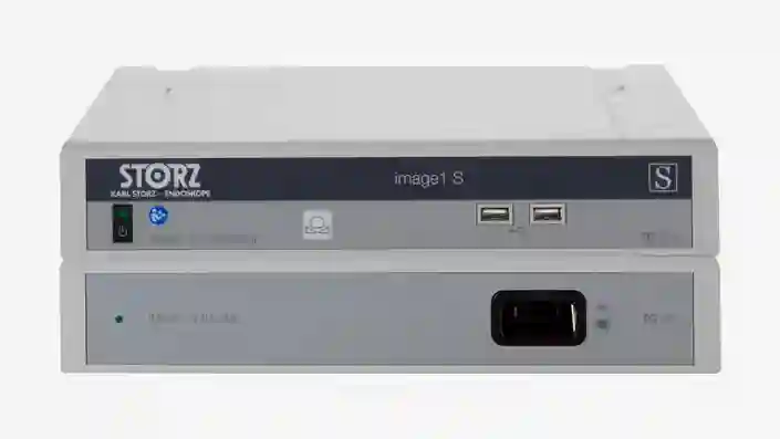
IMAGE 1 S™
Ti offriamo la soluzione adeguata per la visualizzazione ottimale dei tuoi pazienti e per ottenere i migliori risultati di trattamento possibili. La nostra vasta gamma di sistemi di visualizzazione offre soluzioni modulari, sostenibili e personalizzabili in base alle tue esigenze individuali.
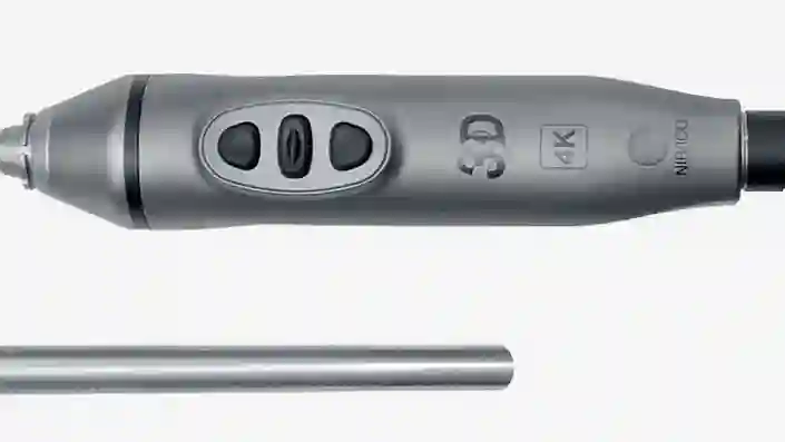
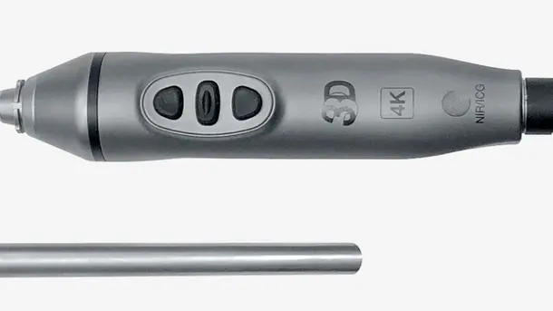
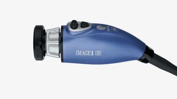
TIPCAM®1 Rubina®
Questo videoendoscopio “all in one” riunisce tre tecnologie di imaging all’avanguardia 4K, 3D e NIR/ICG in un solo prodotto per garantire una migliore visualizzazione e un’eccellente percezione della profondità.
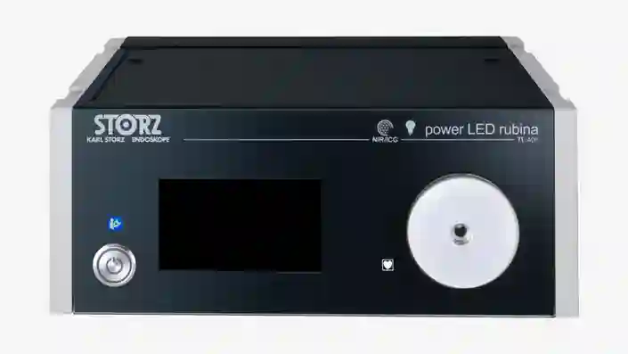
Power LED Rubina®
Questa fonte di luce laser-free è stata sviluppata per applicazioni NIR/ICG e a luce bianca e si contraddistingue per la sua efficienza e lunga durata.
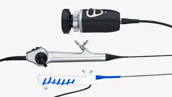
Completa la tua soluzione di imaging NIR/ICG
Configura il sistema con i componenti adatti per rispondere alle tue esigenze individuali.
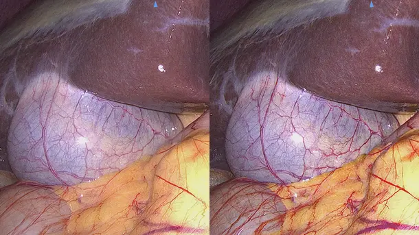
Settori di applicazione IMAGE1 S™ Rubina®
“Il sistema IMAGE1 S™ Rubina® ci ha consentito di fare un grande passo in avanti nel campo della chirurgia assistita tramite la fluorescenza. Siamo usciti “dall’oscurità” e passati alla luce in formato 4K/3D che ci consente di assistere ancor meglio i nostri pazienti.”

IMAGE1 S™ 4U
Con l'introduzione del 4K, che ha rappresentato un passo avanti nello sviluppo della qualità dell'immagine endoscopica, siete in grado di riconoscere e identificare i dettagli più fini durante l'intervento chirurgico. Abbiamo sempre come obiettivo l'ottimizzazione continua della qualità dell'immagine.
Modalità di visualizzazione
Scopri diverse modalità di visualizzazione per una migliore differenziazione e identificazione delle strutture.
CLARA
Le S-Technology assicurano un’illuminazione omogenea, evitando sovraesposizioni e riflessi e, nello stesso tempo, permettono uno schiarimento delle aeree più in ombra.
CHROMA
Con questa modalità di visualizzazione il contrasto cromatico è esaltato senza alterare la naturale percezione del colore. Le differenze di colore e delle strutture vengono quindi visualizzate più chiaramente.
CLARA + CHROMA
La combinazione di queste due S-Technology consente di ottenere una luminosità dell’immagine omogenea e di creare una linea di demarcazione evidente tra le diverse strutture tissutali.
SPECTRA A
Con questa modalità, lo spostamento del colore spettrale nella tonalità del rosso aiuta la differenziazione dei tessuti mostrando delicate strutture rosse come i vasi sanguigni e le mucose nei toni del blu-verde.
SPECTRA B
SPECTRA B riduce le tonalità del rosso e intensifica la porzione di spettro verde-blu grazie alla distorsione colorimetrica. Lo sfondo dell’immagine assume un colore verdognolo, in modo da evidenziare i vasi sanguigni e i capillari.
La famiglia di prodotti IMAGE1 S™ 4U
IMAGE 1 S™
Ti offriamo la soluzione adeguata per la visualizzazione ottimale dei tuoi pazienti e per ottenere i migliori risultati di trattamento possibili. La nostra vasta gamma di sistemi di visualizzazione offre soluzioni modulari, sostenibili e personalizzabili in base alle tue esigenze individuali.
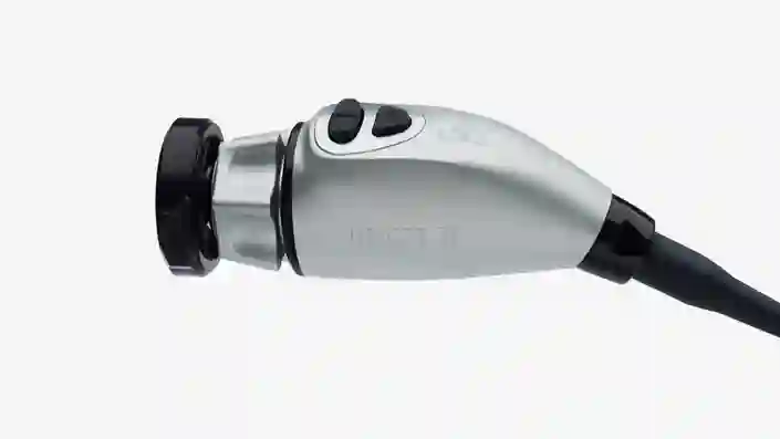
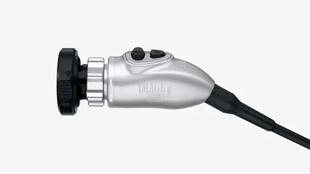
Testina telecamera IMAGE1 S™ 4U
Questa testina rappresenta l’elemento chiave per la visualizzazione. La visualizzazione in 4K ti consente di avere a disposizione una risoluzione più elevata e uno spettro cromatico più ampio per poter identificare e differenziare meglio le strutture tissutali.
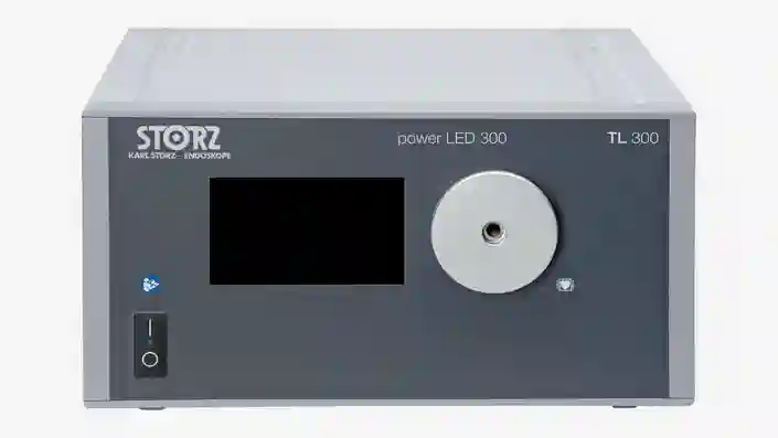
POWER LED 300
Questa fonte di luce laser-free è stata sviluppata per applicazioni a luce bianca e si contraddistingue per la sua efficienza e la sua lunga durata.
Un sistema di immagini in 4K
Combina questi componenti con un sistema adatto alle tue esigenze.
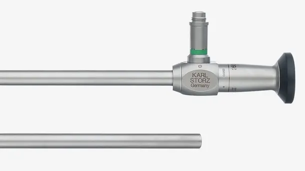
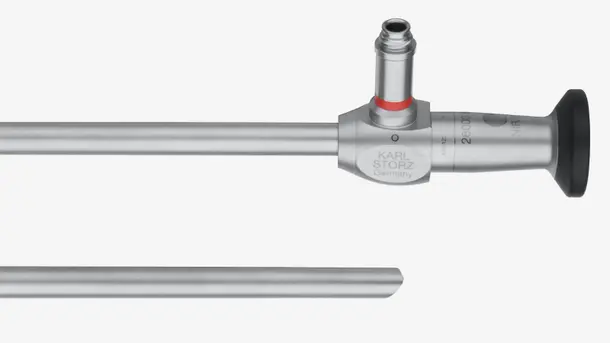
Sistemi ottici HOPKINS®
Scopri le nostre ottiche adatte per applicazioni a luce bianca e compatibili con i nostri sistemi e completa la tua catena di immagini.
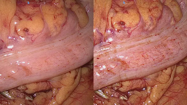
Settori di applicazione IMAGE1 S™ 4U
"Con l’introduzione della tecnologia IMAGE1 S™ nel nostro ospedale, abbiamo visualizzato elementi che non avevamo visto prima. Questo ci ha permesso una migliore visualizzazione per poter eseguire trattamenti più precisi."

IMAGE1 S™ Saphira™
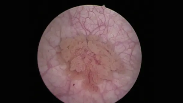
La visualizzazione Blue Light Imaging (BLI), finora conosciuta col nome di PDD, viene utilizzata dopo la somministrazione del farmaco Hexvix/Cysview e consente la visualizzazione di tumori maligni allo stadio precoce che spesso non riescono ad essere riconosciuti nella modalità a luce bianca.
Modalità di visualizzazione
Scopri diverse modalità di visualizzazione per una migliore differenziazione e identificazione delle strutture.
BLI
La luce blu rende più agevole la distinzione e l’identificazione delle cellule tumorali che appaiono in rosso fluorescente rispetto ai tessuti adiacenti che sono di colore blu.
CHROMA
Con questa modalità di visualizzazione il contrasto cromatico è esaltato senza alterare la naturale percezione del colore. Le differenze di colore e delle strutture vengono quindi visualizzate più chiaramente.
SPECTRA A
Con questa modalità, lo spostamento del colore spettrale nella tonalità del rosso aiuta la differenziazione dei tessuti mostrando delicate strutture rosse come i vasi sanguigni e le mucose nei toni del blu-verde.
SPECTRA B
SPECTRA B riduce le tonalità del rosso e intensifica la porzione di spettro verde-blu grazie alla distorsione colorimetrica. Lo sfondo dell’immagine assume un colore verdognolo, in modo da evidenziare i vasi sanguigni e i capillari.
La famiglia di prodotti IMAGE1 S™ Saphira™
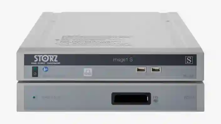
IMAGE 1 S™
Ti offriamo la soluzione adeguata per la visualizzazione ottimale dei tuoi pazienti e per ottenere i migliori risultati di trattamento possibili. La nostra vasta gamma di sistemi di visualizzazione offre soluzioni modulari, sostenibili e personalizzabili in base alle tue esigenze individuali.
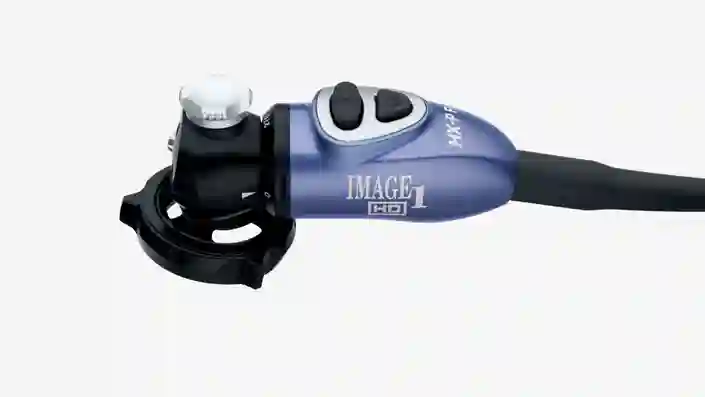
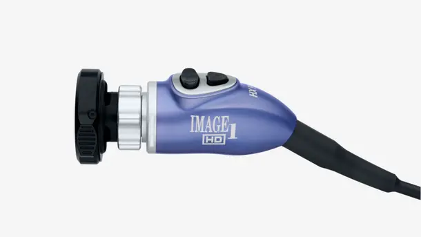
Testina IMAGE1 S™ HX-P FI
Questa testina pendulum è stata sviluppata per applicazioni Blue Light Imaging e a luce bianca. Il suo design e il suo peso ridotto ti consentono un lavoro ergonomico.

POWER LED Saphira™
Questa fonte di luce laser-free è stata sviluppata per applicazioni Blue Light Imaging e a luce bianca e si contraddistingue per la sua efficienza e lunga durata.
Completa la tua soluzione Blue Light Imaging
Configura il sistema con i componenti adatti per rispondere alle tue esigenze individuali.
Sistemi ottici HOPKINS® BLI
Scopri le nostre ottiche ottimizzate per applicazioni Blue Light Imaging e a luce bianca, compatibili con i nostri sistemi e completa la tua catena di immagini.
Campi di applicazione IMAGE1 S™ Saphira™
“Ho avuto occasione negli ultimi mesi di utilizzare la nuova sorgente luminosa Saphira™ e posso dire che il suo uso ha contribuito a migliorare notevolmente la qualità delle procedure.”

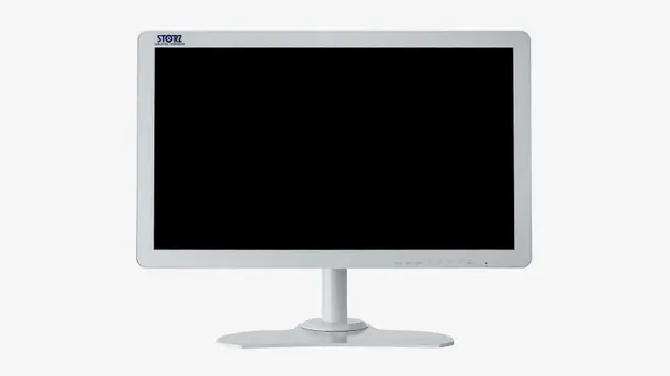
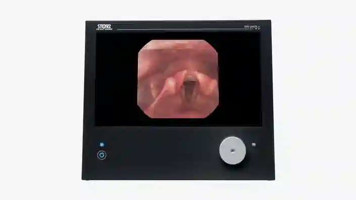
TELE PACK+
Questa piattaforma compatta è stata sviluppata per la diagnosi e per piccoli interventi. Offre molte opzioni di compatibilità e questo la rende ideale per l’impiego in studi medici, day hospital, Pronto Soccorso, reparti di medicina intensiva ed in ambito ambulatoriale.
La famiglia di prodotti TELE PACK+
TELE PACK+
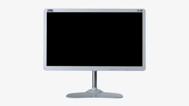
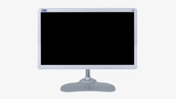
Sperimenta i vantaggi di un’apparecchiatura compatta e portatile che riunisce in sè il monitor, la fonte di luce LED, l’unità controllo di telecamera e la documentazione con funzione di rete.

X-Line e C-Line
Grazie ai suoi attacchi X-Line e C-Line, TELE PACK+ è compatibile con numerosi endoscopi rigidi, flessibili e monouso ed è quindi adatta per l’impiego in quasi tutte le discipline medicali.
Completa la tua soluzione compatta di imaging FULL HD
Combina questi componenti con un sistema adatto alle tue esigenze
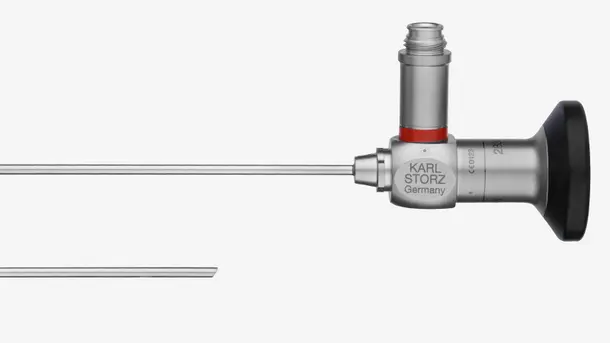
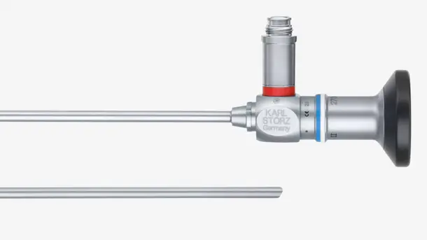
Endoscopi rigidi
Scopri le nostre ottiche rigide adatte per applicazioni a luce bianca e compatibili con i nostri sistemi e completa la tua catena di immagini.
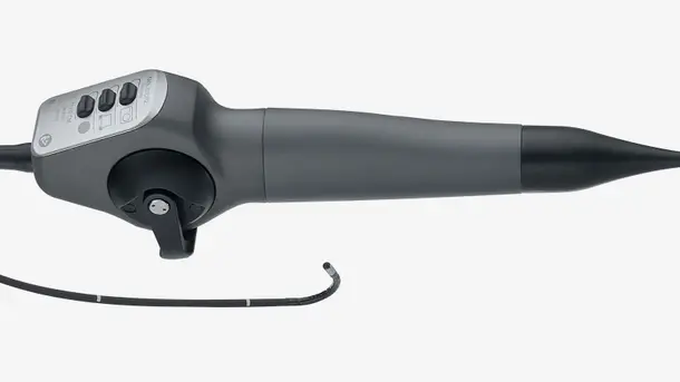
Videoendoscopi
Scopri la nostra vasta scelta di videoendoscopi flessibili che ti aiuteranno a visualizzare cavità strette o difficili da raggiungere, evitando interventi invasivi. Il nostro ampio portfolio di videoendoscopi compatibili ti offrono una soluzione per quasi tutte le discipline medicali.
Campi di applicazione TELE PACK+
"Apprezziamo molto il nuovo sistema TELE PACK+, perchè fornisce una qualità brillante di immagini che consente di formulare diagnosi precise anche in casi difficili."

TELECAM C3
La TELECAM C3 è ideale per l’esecuzione di piccole procedure diagnostiche sia in studi medici che in altri ambienti chirurgici. Questa unità è stata sviluppata per l’impiego con un'ampia gamma di endoscopi compatibili e può essere utilizzata in quasi tutte le discipline medicali.
La famiglia di prodotti TELECAM C3
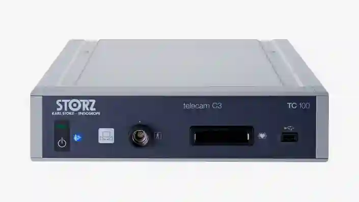
TELECAM C3
La nostra unità di controllo telecamera economica e compatta è stata sviluppata per eseguire interventi endoscopici semplici. Dispone di due attacchi per telecamera che consentono la compatibilità con un gran numero di endoscopi.
X-Line e C-Line
Grazie ai suoi attacchi X-Line e C-Line, TELECAM C3 è compatibile con numerosi endoscopi rigidi, flessibili e monouso e quindi adatta per l’impiego in quasi tutte le discipline medicali.
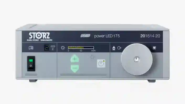
POWER LED 175
Questa fonte di luce laser-free è stata sviluppata per applicazioni a luce bianca nelle cavità più piccole e si contraddistingue per la sua efficienza e lunga durata.
Completa la tua soluzione di imaging FULL HD
Combina questi componenti con un sistema adatto alle tue esigenze.
Endoscopi rigidi
Scopri le nostre ottiche rigide adatte per applicazioni a luce bianca e compatibili con i nostri sistemi e completa la tua catena di immagini.
Videoendoscopi
Scopri la nostra vasta scelta di videoendoscopi flessibili che ti aiuteranno a visualizzare cavità strette o difficili da raggiungere, evitando interventi invasivi. Il nostro ampio portfolio di videoendoscopi compatibili ti offrono una soluzione per quasi tutte le discipline medicali.
Settori di applicazione di TELECAM C3
Siamo in grado di creare sale operatorie all’avanguardia personalizzandole in base alle tue esigenze.
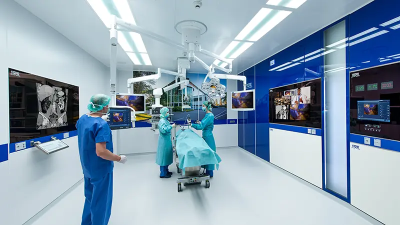
La sala operatoria del futuro
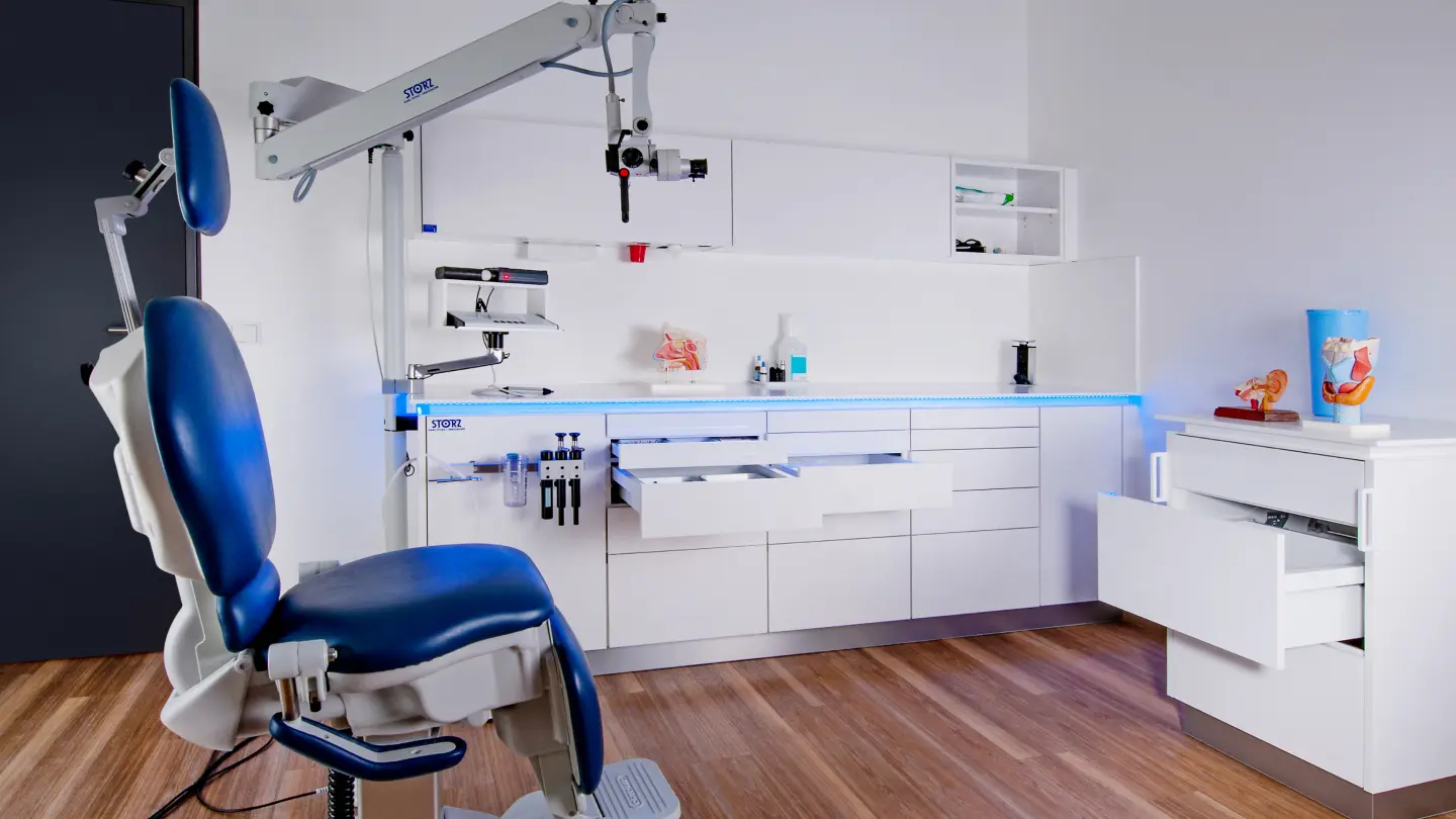
Soluzione di arredo modulare per l’ambulatorio
Sapere è potere
Siamo consapevoli come oggigiorno le tecniche chirurgiche, gli strumenti e le apparecchiature utilizzate diventino sempre più complessi ed aumentino quindi la necessità e l’importanza di una formazione professionale mirata, di corsi di aggiornamento adeguati che consentano un corretto sviluppo professionale. Mettiamo a disposizione dei medici e degli operatori del settore di tutto il mondo, i nostri corsi di aggiornamento, workshop e congressi personalizzati.


CONTATTO Contatta il nostro specialista.
Ti guidiamo lungo l’intero processo e ti aiutiamo a trovare la soluzione che più si adatta alle tue esigenze in chirurgia.