
Bildgebung
Als Pionier in der endoskopischen Bildgebung bietet KARL STORZ Ihnen innovative, skalierbare und modulare Lösungen, die Sie dabei unterstützen, die optimale Bildkette für Ihre Bedürfnisse und Ihr Budget zusammenzustellen.
Als Pionier in der endoskopischen Bildgebung bietet KARL STORZ Ihnen innovative, skalierbare und modulare Lösungen, die Sie dabei unterstützen, die optimale Bildkette für Ihre Bedürfnisse und Ihr Budget zusammenzustellen.
Vorteile
Präzise. Modular. Skalierbar.
.svg)


Produkte im Spotlight


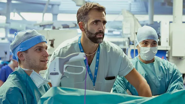
Vielseitige Bildgebung
Unsere Bildgebungstechnologien ermöglichen den Zugang zu optimierten Visualisierungsmöglichkeiten im OP und in der Praxis. Mit unterschiedlichen Konfigurationsoptionen können Sie eine Bildkette zusammenstellen, die Ihren Bedürfnissen und Ihrem Budget am besten entspricht.
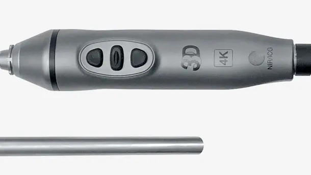
IMAGE1 S™ Rubina®
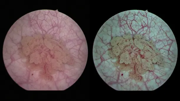
Durch die Kombination von 3D- und 4K-Technologie mit NIR/ICG-Fluoreszenzbildgebung werden qualitativ hochwertige Informationen bereitgestellt, um die Detektion und die chirurgische Präzision zu verbessern. NIR/ICG erweitert die diagnostischen Möglichkeiten, z. B. die Perfusionsbeurteilung und die Detektion der Sentinel-Lymphknoten.
Visualisierungsmodi
Entdecken Sie verschiedene Visualisierungsmodi, die Sie dabei unterstützen, Strukturen differenzierter zu erkennen.
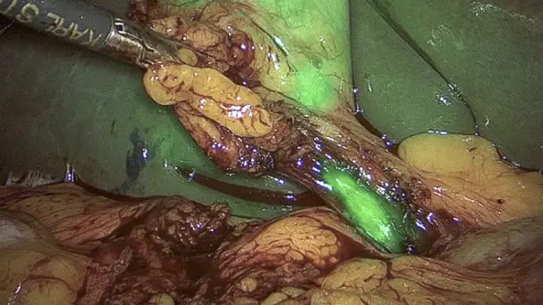
Overlay
Dieser Modus kombiniert das reguläre Weißbild mit NIR/ICG-Informationen, um ein grünes oder blaues Überlagerungsbild zu genereieren.
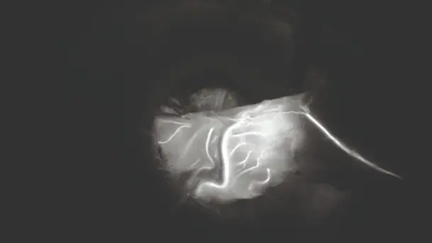
Monochromatisch
Das NIR/ICG-Signal wird in Weiß auf schwarzem Hintergrund dargestellt, um eine bestmögliche Differenzierung zu erreichen.
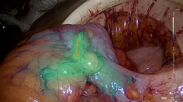
Intensity Map
Die Intensität des NIR/ICG-Signals wird mittels Farbskala in einem Überlagerungsbild dargestellt.
CLARA
Diese S-Technologie sorgt für eine homogene Ausleuchtung, verhindert Überbelichtung und Reflexionen und verbessert die Sichtbarkeit in dunklen Bereichen.
CHROMA
Diese S-Technologie intensiviert den Farbkontrast, ohne die natürliche Farbwahrnehmung zu verändern. Farbunterschiede und Strukturen werden dadurch prägnanter dargestellt.
CLARA + CHROMA
Die Kombination dieser beiden S-Technologien ermöglicht eine homogene Ausleuchtung des Bilds und die Hervorhebung von Gewebestrukturen.
SPECTRA A
Diese S-Technologie unterstützt durch die spektrale Farbtonverschiebung die Gewebedifferenzierung, indem feine rote Strukturen wie Blutgefäße und Schleimhäute in grünlich-blauem Licht dargestellt werden.
SPECTRA B
Durch die spektrale Farbtonverschiebung werden Rottöne reduziert und die grünlich-blauen Spektralanteile verstärkt. Der Hintergrund wirkt dadurch grünlich und Blutgefäße und Kapillaren werden hervorgehoben.
Die IMAGE1 S™ Rubina® Produktfamilie
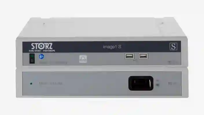
IMAGE 1 S™
Wir bieten Ihnen die passende Lösung für die optimale Visualisierung Ihrer Patientinnen und Patienten, damit Sie das bestmögliche Behandlungsergebnis erreichen. Mit unseren modularen, nachhaltigen und individuell auf Ihre Bedürfnisse anpassbaren Lösungen stellen wir eine breite Palette an Visualisierungsmöglichkeiten bereit.
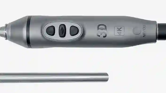
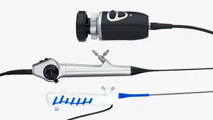
TIPCAM®1 Rubina®
Dieses All-in-one-Videoendoskop vereint drei fortschrittliche Bildgebungstechnologien – 4K, 3D und NIR/ICG – in einem einzigen Produkt für eine verbesserte Visualisierung und eine optimale Tiefenwahrnehmung.
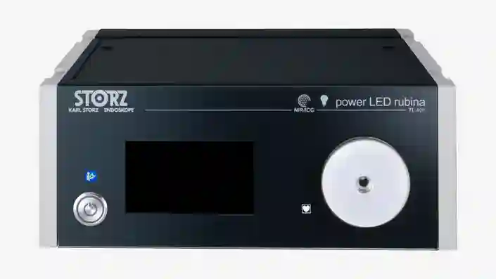
Power LED Rubina®
Diese laserfreie Kaltlichtquelle wurde für Weißlicht- und NIR/ICG-Anwendungen entwickelt und zeichnet sich durch Effizienz und Langlebigkeit aus.
Vervollständigen Sie Ihre NIR/ICG-Bildgebung
Kombinieren Sie diese Komponenten zu einem an Ihre Bedürfnisse angepassten System.
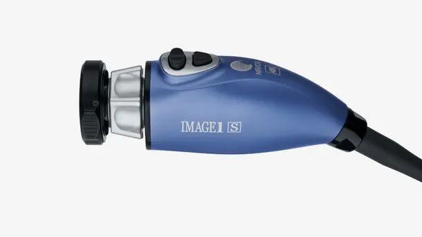
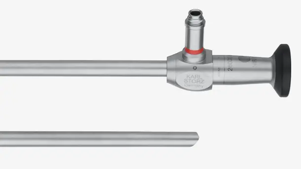
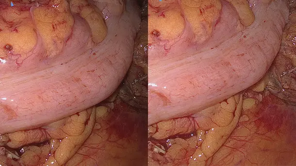
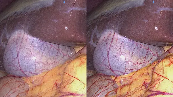
IMAGE1 S™ Rubina® Anwendungsbereiche
Dank des IMAGE1 S™ Rubina® Systems haben wir in der fluoreszenzgestützten Chirurgie einen großen Schritt nach vorn gemacht. Wir sind aus der „Dunkelheit“ ins 4K/3D-Licht getreten und können unsere Patienten nun noch besser versorgen.

IMAGE1 S™ 4U
Die Einführung von 4K – der nächsten Stufe in der Entwicklung der endoskopischen Bildqualität – versetzt Sie in die Lage, während der Operation feine Details zu erkennen und zu identifizieren, und bringt uns auf unserem Weg der kontinuierlichen Optimierung der Bildgebung einen Schritt weiter.
Visualisierungsmodi
Entdecken Sie verschiedene Visualisierungsmodi, die Sie dabei unterstützen, Strukturen differenzierter zu erkennen.
CLARA
Diese S-Technologie sorgt für eine homogene Ausleuchtung, verhindert Überbelichtung und Reflexionen und verbessert die Sichtbarkeit in dunklen Bereichen.
CHROMA
Diese S-Technologie intensiviert den Farbkontrast, ohne die natürliche Farbwahrnehmung zu verändern. Farbunterschiede und Strukturen werden dadurch prägnanter dargestellt.
CLARA + CHROMA
Die Kombination dieser beiden S-Technologien ermöglicht eine homogene Ausleuchtung des Bilds und die Hervorhebung von Gewebestrukturen.
SPECTRA A
Diese S-Technologie unterstützt durch die spektrale Farbtonverschiebung die Gewebedifferenzierung, indem feine rote Strukturen wie Blutgefäße und Schleimhäute in grünlich-blauem Licht dargestellt werden.
SPECTRA B
Durch die spektrale Farbtonverschiebung werden Rottöne reduziert und die grünlich-blauen Spektralanteile verstärkt. Der Hintergrund wirkt dadurch grünlich und Blutgefäße und Kapillaren werden hervorgehoben.
Die IMAGE1 S™ 4U Produktfamilie
IMAGE 1 S™
Wir bieten Ihnen die passende Lösung für die optimale Visualisierung Ihrer Patientinnen und Patienten, damit Sie das bestmögliche Behandlungsergebnis erreichen. Mit unseren modularen, nachhaltigen und individuell auf Ihre Bedürfnisse anpassbaren Lösungen stellen wir eine breite Palette an Visualisierungsmöglichkeiten bereit.
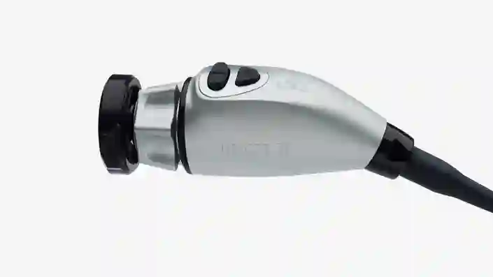
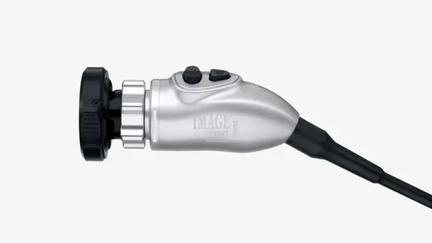
IMAGE1 S™ 4U Kamerakopf
Dieser Kamerakopf ist ein Schlüsselelement für die Visualisierung. Mit seiner 4K-Darstellung profitieren Sie von einer höheren Auflösung und einem erweiterten Farbraum und werden so bei der Identifizierung und Differenzierung von Gewebestrukturen unterstützt.
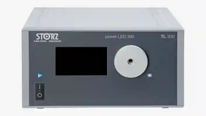
POWER LED 300
Diese laserfreie Kaltlichtquelle wurde für Weißlichtanwendungen entwickelt und zeichnet sich durch Effizienz und Langlebigkeit aus.
Vervollständigen Sie Ihre 4K-Bildgebung
Kombinieren Sie diese Komponenten zu einem an Ihre Bedürfnisse angepassten System.
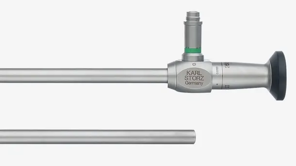
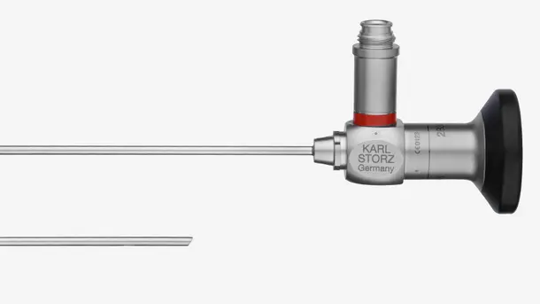
HOPKINS® Optiken
Entdecken Sie unsere systemkompatiblen, für Weißlichtanwendungen optimierten Optiken und vervollständigen Sie Ihre Bildgebungskette.
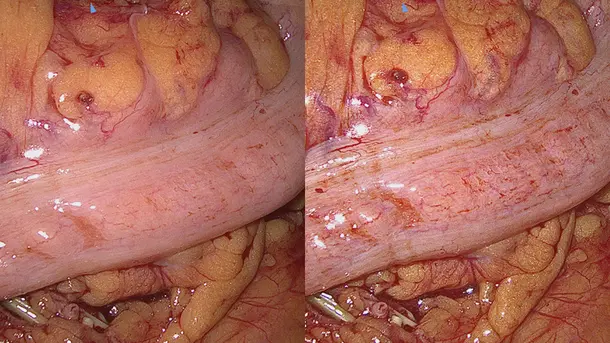
IMAGE1 S™ 4U Anwendungsbereiche
Als die IMAGE1 S™ Technologie in unserem Krankenhaus eingeführt wurde, haben wir plötzlich Dinge gesehen, die wir vorher nie gesehen hatten. Wenn man besser sieht, kann man besser behandeln.

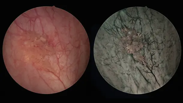
IMAGE1 S™ Saphira™
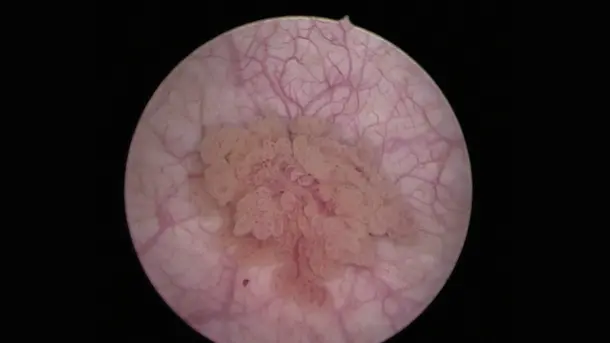
Blue Light Imaging (BLI), bisher auch als PDD bekannt, ermöglicht bei Verabreichung von Hexvix/Cysview die Visualisierung von malignen Tumoren im Frühstadium, die in der Weißlicht-Diagnostik oftmals nicht oder nur schwer erkennbar sind.
Visualisierungsmodi
Entdecken Sie verschiedene Visualisierungsmodi, die Sie dabei unterstützen, Strukturen differenzierter zu erkennen.
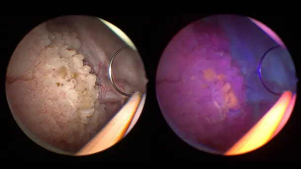
BLI
Durch die Beleuchtung mit blauem Licht wird die Unterscheidung der rot fluoreszierenden Tumorzellen vom umgebenden Gewebe, das in blauer Farbe dargestellt wird, erleichtert.
CHROMA
Diese S-Technologie intensiviert den Farbkontrast, ohne die natürliche Farbwahrnehmung zu verändern. Farbunterschiede und Strukturen werden dadurch prägnanter dargestellt.
SPECTRA A
Diese S-Technologie unterstützt durch die spektrale Farbtonverschiebung die Gewebedifferenzierung, indem feine rote Strukturen wie Blutgefäße und Schleimhäute in grünlich-blauem Licht dargestellt werden.
SPECTRA B
Durch die spektrale Farbtonverschiebung werden Rottöne reduziert und die grünlich-blauen Spektralanteile verstärkt. Der Hintergrund wirkt dadurch grünlich und Blutgefäße und Kapillaren werden hervorgehoben.
Die IMAGE1 S™ Saphira™ Produktfamilie
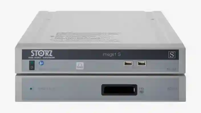
IMAGE 1 S™
Wir bieten Ihnen die passende Lösung für die optimale Visualisierung Ihrer Patientinnen und Patienten, damit Sie das bestmögliche Behandlungsergebnis erreichen. Mit unseren modularen, nachhaltigen und individuell auf Ihre Bedürfnisse anpassbaren Lösungen stellen wir eine breite Palette an Visualisierungsmöglichkeiten bereit.
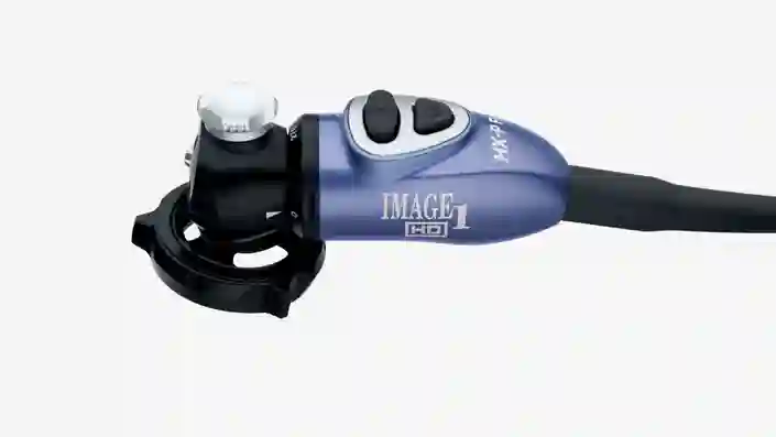
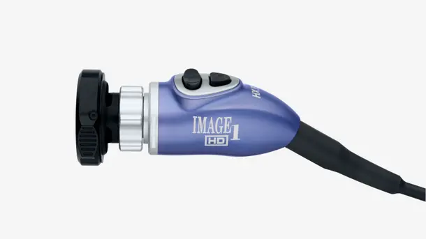
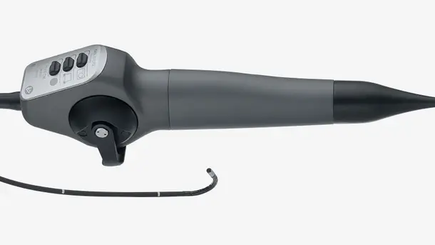
IMAGE1 S™ HX-P FI-Camera Head
Dieser Pendelkamerakopf wurde für Blue Light- und Weißlichtanwendungen entwickelt und ermöglicht durch seine Bauweise und sein geringes Gewicht ergonomisches Arbeiten.

POWER LED Saphira™
Diese laserfreie Kaltlichtquelle wurde für Weißlichtanwendungen und Blue Light Imaging entwickelt und zeichnet sich durch Effizienz und Langlebigkeit aus.
Vervollständigen Sie Ihre Blue-Light-Imaging-Lösung
Kombinieren Sie diese Komponenten zu einem an Ihre Bedürfnisse angepassten System.
HOPKINS® BLI-Optiken
Entdecken Sie unsere systemkompatiblen, für Blue-Light- und Weißlichtanwendungen optimierten Optiken und vervollständigen Sie Ihre Bildkette.
IMAGE1 S™ Saphira™ Anwendungsbereiche
In den letzten Monaten hatte ich die Gelegenheit, die neue Saphira™-Lichtquelle zu verwenden, die die Qualität des Verfahrens wirklich erheblich verbessert.

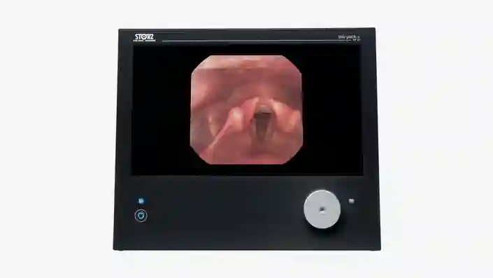
TELE PACK+
Diese kompakte Plattform für die Diagnose und kleinere Eingriffe eignet sich dank zahlreicher Kompatibilitätsoptionen ideal für den Einsatz in Arztpraxen, Tageskliniken, Notfallstationen, Intensivstationen und Ambulanzen.
Die TELE PACK+ Produktfamilie
TELE PACK+
Erleben Sie die Vorteile eines kompakten und portablen Geräts, das Monitor, LED-Lichtquelle, Kamerakontrolleinheit und Dokumentation mit integrierter Netzwerkfunktion vereint.

X-Line und C-Line
Mit seinen X-Line- und C-Line-Anschlüssen ist der TELE PACK+ mit einer Vielzahl an starren, flexiblen und Einweg-Endoskopen kompatibel und somit für den Einsatz in nahezu allen medizinischen Fachgebieten geeignet.
Vervollständigen Sie Ihre kompakte FULL HD-Bildgebung
Kombinieren Sie diese Komponenten zu einem an Ihre Bedürfnisse angepassten System.
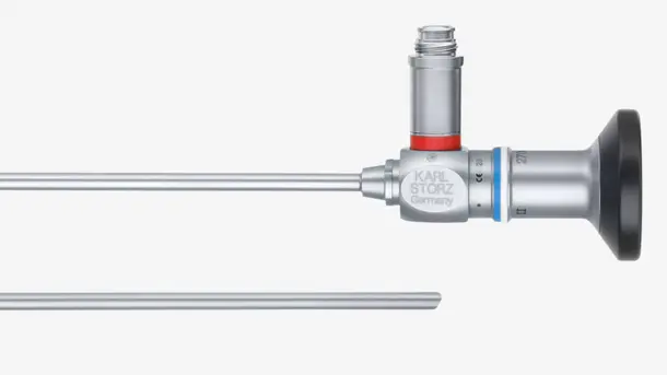
Starre Endoskope
Entdecken Sie unsere systemkompatiblen, für Weißlichtanwendungen optimierten starren Optiken und vervollständigen Sie Ihre Bildgebungskette.
Videoendoskope
Entdecken Sie unser umfangreiches Angebot an flexiblen Videoendoskopen, mit denen enge oder verwinkelte Kavitäten ohne invasive Eingriffe sichtbar gemacht werden können. Mit einer Vielzahl an kompatiblen Videoendoskopen bieten wir Ihnen eine Lösung für nahezu alle Fachgebiete.
TELE PACK+ Anwendungsbereiche
Das neue TELE PACK+ System gefällt uns sehr gut, weil es über eine brillante Bildqualität verfügt, die auch bei schwierigen Fällen eine effektive Diagnose ermöglicht.

TELECAM C3
Die Kamerakontrolleinheit TELECAM C3 eignet sich optimal für die Diagnose und kleinere Eingriffe in Arztpraxen und im chirurgischen Umfeld. Ausgelegt für die Verwendung mit einer Vielzahl an kompatiblen Endoskopen kann sie in nahezu allen Fachgebieten eingesetzt werden.
Die TELECAM C3 Produktfamilie
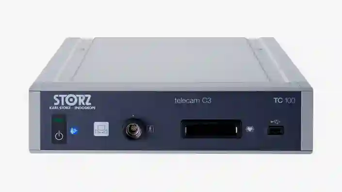
TELECAM C3
Unsere kostengünstige und kompakte Kamerakontrolleinheit wurde für einfache endoskopische Eingriffe entwickelt und verfügt über zwei Kameraanschlüsse, die die Kompatibilität mit einer Vielzahl von Endoskopen sicherstellen.
X-Line and C-Line
Mit seinen X-Line- und C-Line-Anschlüssen ist die TELECAM C3 mit einer Vielzahl an starren, flexiblen und Einweg-Endoskopen kompatibel und somit für den Einsatz in nahezu allen medizinischen Fachgebieten geeignet.
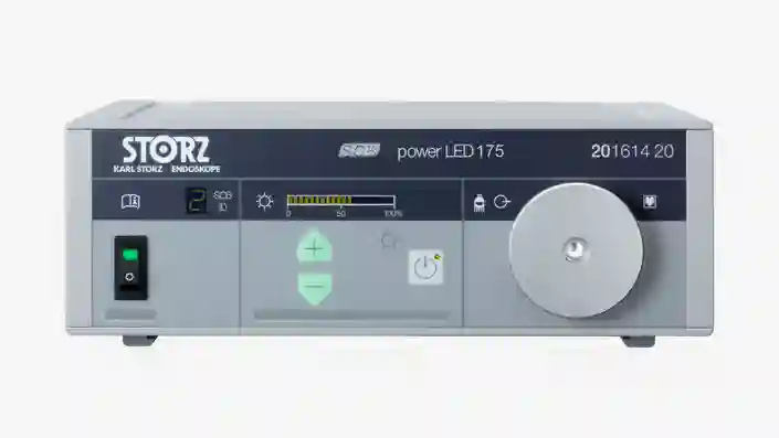
POWER LED 175
Diese laserfreie Kaltlichtquelle wurde für Weißlichtanwendungen in kleineren Kavitäten entwickelt und zeichnet sich durch Effizienz und Langlebigkeit aus.
Vervollständigen Sie Ihre FULL HD-Bildgebung
Kombinieren Sie diese Komponenten zu einem an Ihre Bedürfnisse angepassten System.
Starre Endoskope
Entdecken Sie unsere systemkompatiblen, für Weißlichtanwendungen optimierten starren Optiken und vervollständigen Sie Ihre Bildgebungskette.
Videoendoskope
Entdecken Sie unser umfangreiches Angebot an flexiblen Videoendoskopen, mit denen enge oder verwinkelte Kavitäten ohne invasive Eingriffe sichtbar gemacht werden können. Mit einer Vielzahl an kompatiblen Videoendoskopen bieten wir Ihnen eine Lösung für nahezu alle Fachgebiete.
TELECAM C3 Anwendungsbereiche
Gestalten Sie hochmoderne Operationssäle, die individuell auf Ihre Bedürfnisse abgestimmt sind
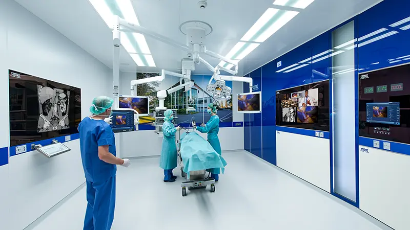
Die Zukunft der Integration im Operationssaal
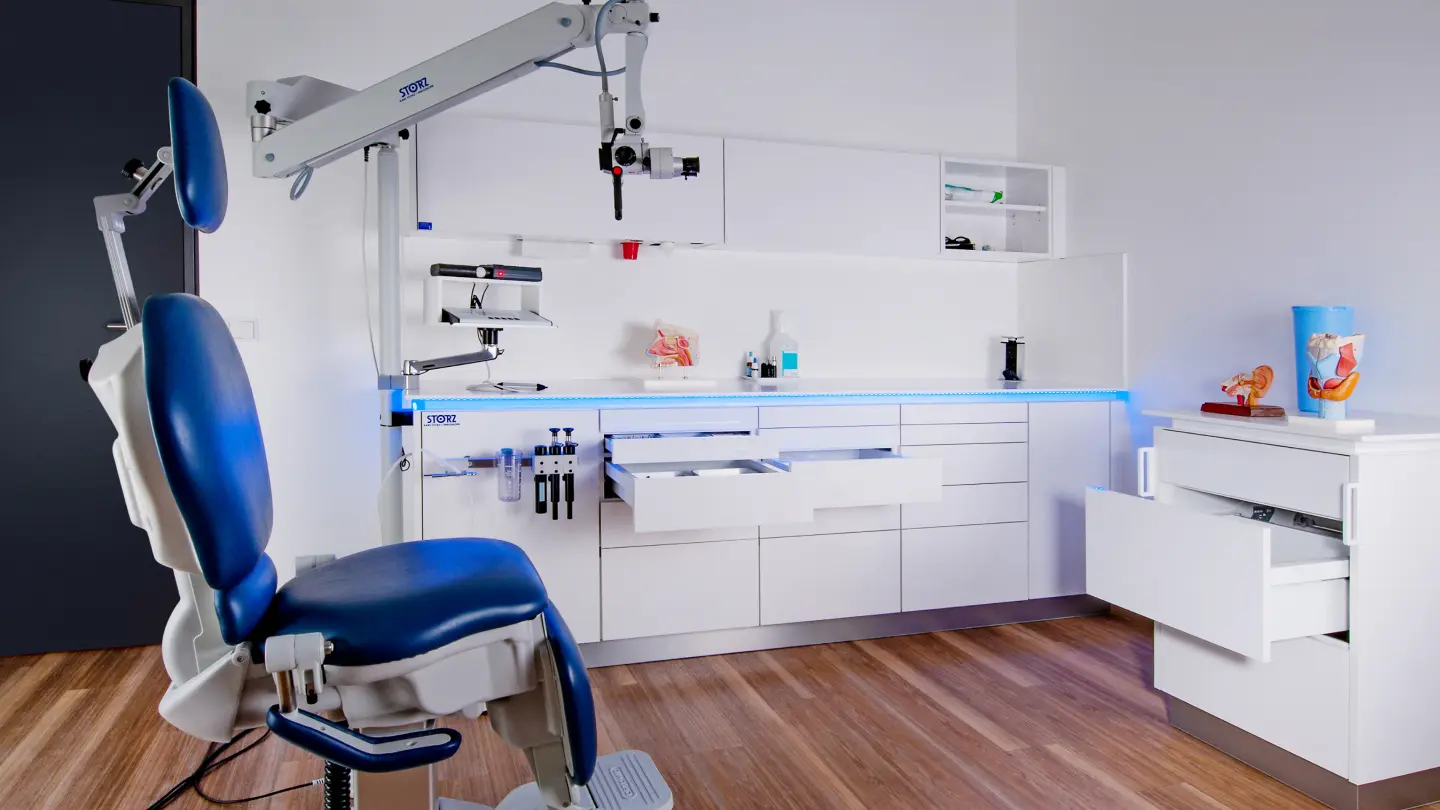
Modulare Innenräume für Ihre Praxis
Wissen ist Macht
Wir sind uns bewusst, dass mit der Komplexität der Chirurgietechniken und der eingesetzten Instrumente und Geräte auch die Bedeutung der zielgerichteten Ausbildung, Weiterbildung und beruflichen Entwicklung zunimmt. Mit maßgeschneiderten Fortbildungskursen, Workshops und Kongressen sind wir bestrebt, Ärzte und Assistenten weltweit zu unterstützen.


Kontakt Nehmen Sie Kontakt mit uns auf
Wir führen Sie durch den gesamten Prozess und helfen Ihnen, die richtige Lösung für Ihre chirurgischen Anforderungen zusammenzustellen.