
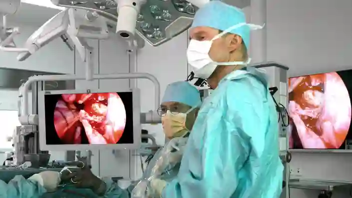
ENT / Otorhinolaryngology
Over 75 years of redefining early ENT diagnosis and interventions by Enabling Anywhere Care.
Over 75 years of redefining early ENT diagnosis and interventions by Enabling Anywhere Care.
BENEFITS
Efficient. Adaptable. Innovative.
.svg)
.svg)

Solutions in the Spotlight
Highlights by Procedure
Ear/Otology
KARL STORZ supports otologists in minimally invasive procedures with power unit, instruments and telescopes. Find out about solutions for ORs and Office clinics as well as innovations in 3D endo/exoscopy in ear and temporal bone surgery.
Everything you’ll need to get started
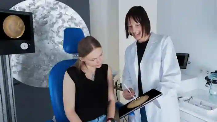
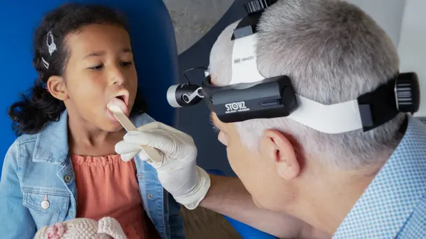
Ear diagnosis
Offering close-up and detailed imaging, illumination, and exposure solutions.
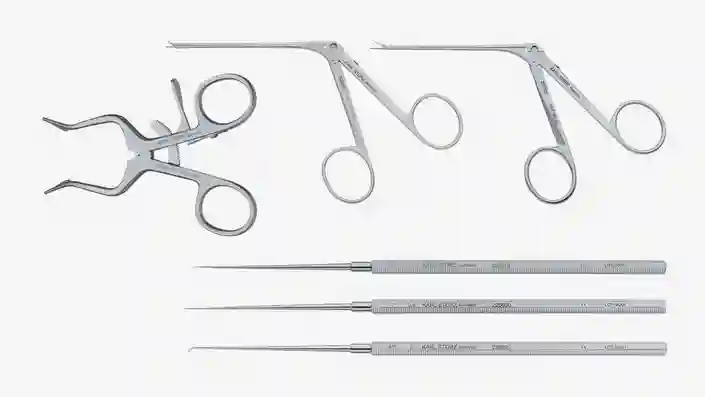
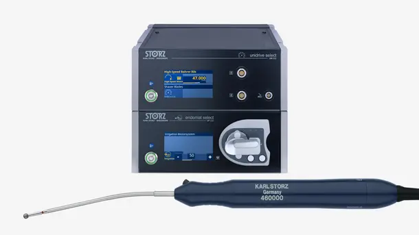
Ear microsurgery
Variety of fine instruments and modern motor system for ear and temporal bone microsurgery.
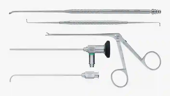
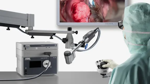
Endoscopic middle ear surgery
Enhance your visualization in middle ear surgery with our 2D and 3D endoscopes.
Highlights from our range


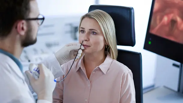
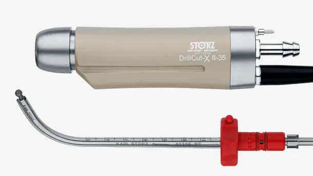
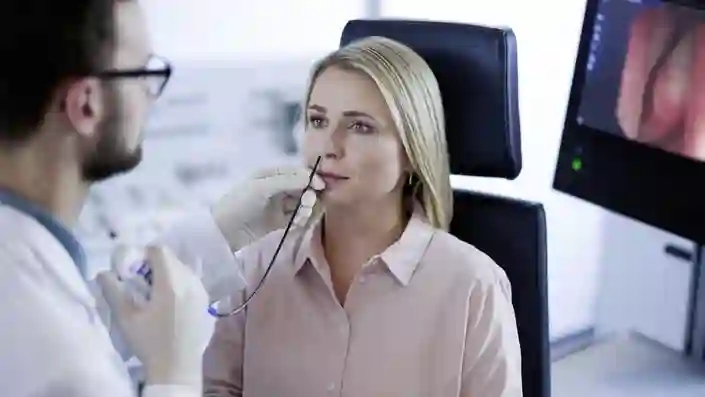
Nose/Rhinology
KARL STORZ offers advanced solutions for rhinologists. We aim to assist physicians in raising the quality of care resulting in better outcomes.
Everything you’ll need to get started
Highlights from our range

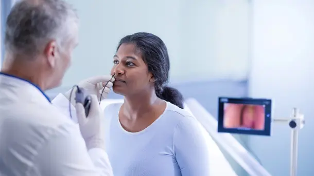
Throat/Laryngology
In the field of laryngology, KARL STORZ has long been a reliable and innovative partner, providing a variety of solutions for customizing ORs and offices.
Everything you’ll need to get started
Office Based Laryngology
Providing instrumentation and compact visualization options, including reusable and single-use videoendoscope options.

OR Based Laryngology
Instrumentation, visualization, and imaging modalities for indications in the mouth, oropharynx, larynx and head and neck.
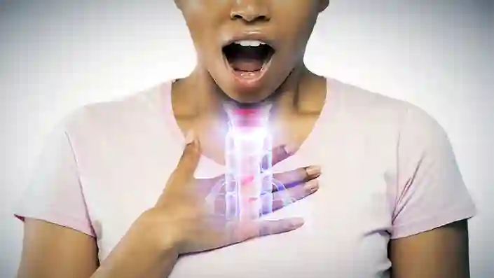
Esophagoscopy
For the treatment of patients, KARL STORZ offers a variety of instrumentation and visualization options.
Highlights from our range

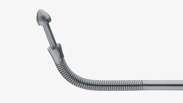
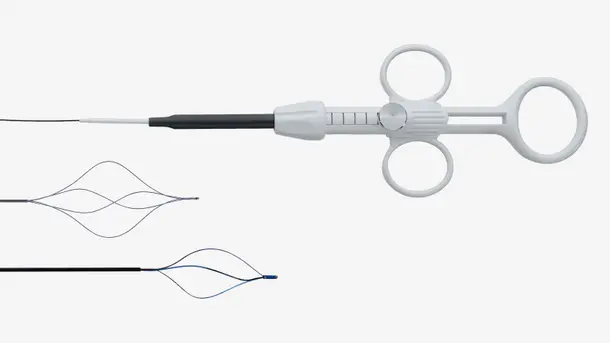
Sialendoscopy
Since more than 20 years, KARL STORZ has been providing innovative solutions in sialendoscopy. Our primary goal is to assist in improving the standard of care, resulting in better outcomes.
Everything you’ll need to get started
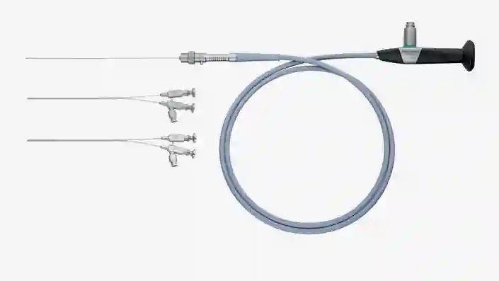
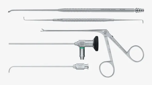
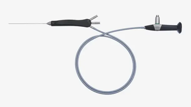
Sialendoscopy
A wide range of miniature autoclavable sialendoscopes and instruments, as well as consumables, are available.
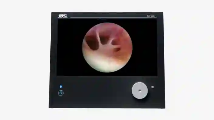
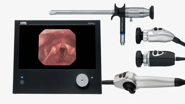
TELE PACK+
Full HD ALL-IN-ONE video system, combining essential components to enable endoscopic diagnosis and treatment.
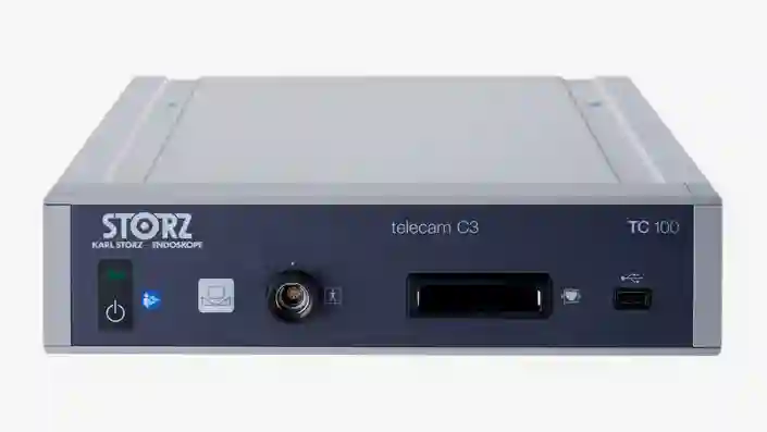
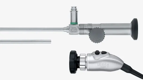
TELECAM C3
Full HD video system, working with sialendoscopes, rigid and flexible endoscopes.
Highlights from our range

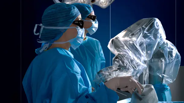
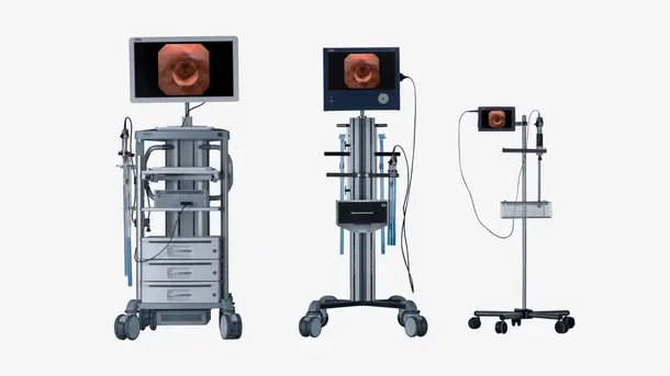
Create state-of-the-art surgical spaces tailored to your needs
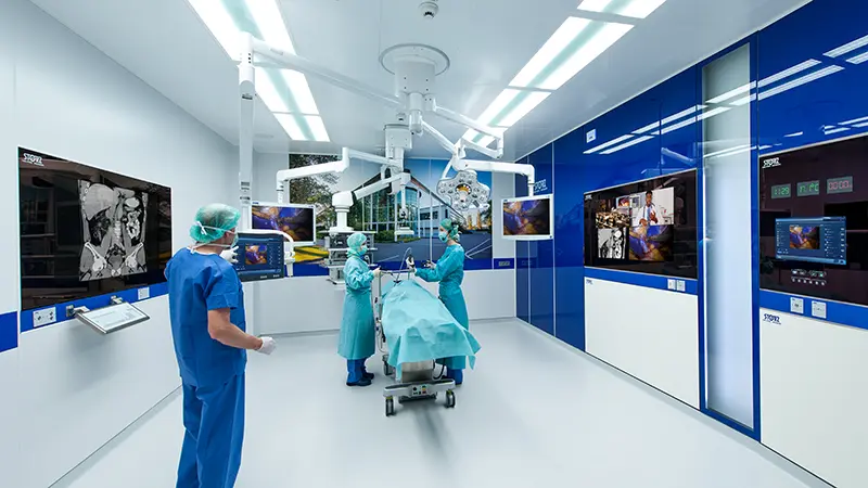
The future of operating room integration
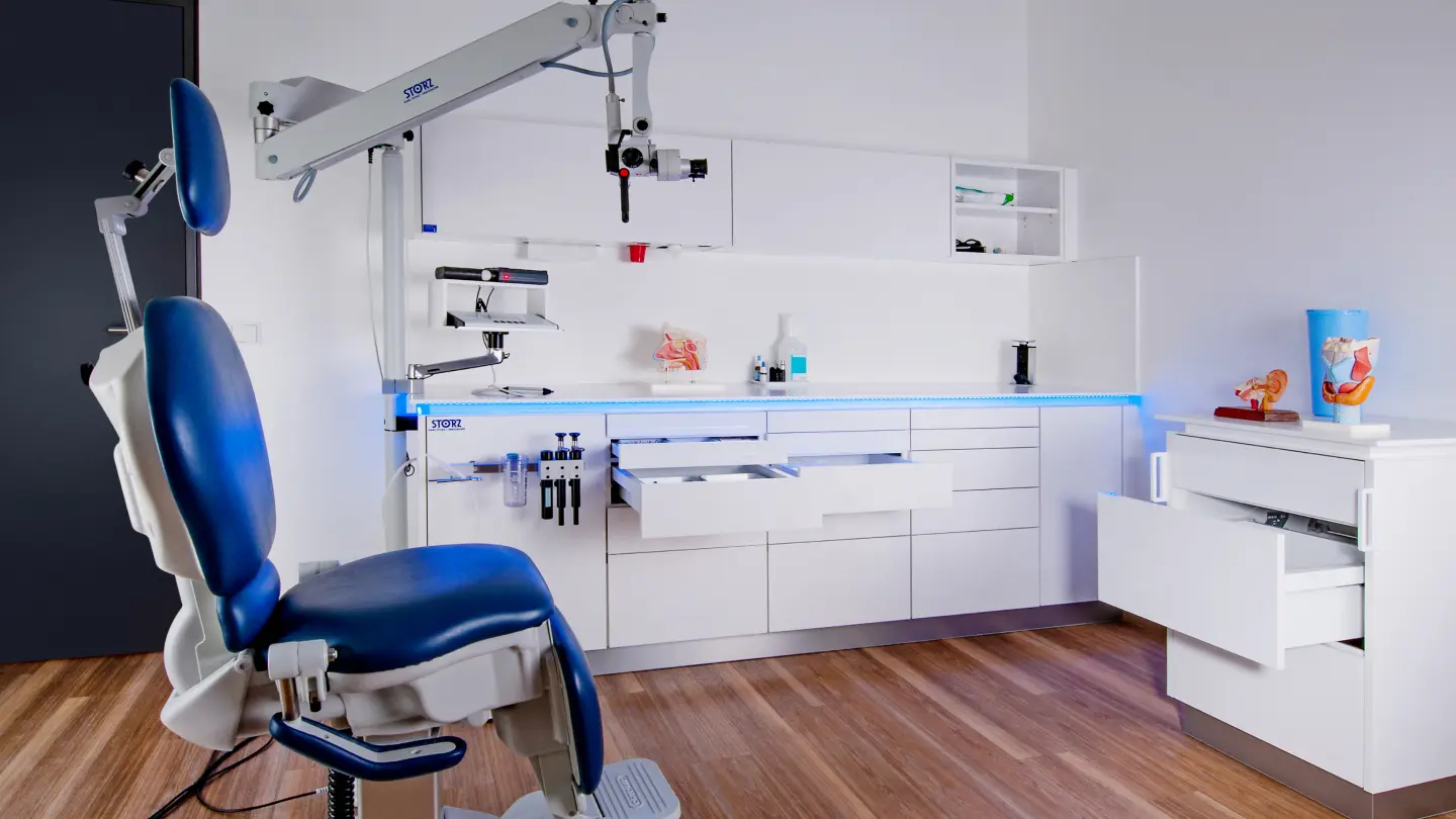
Modular interiors for your practice
Empowering through education
CONTACT Get in touch with your specialist
We’ll guide you through the whole process and help customize the right solution for your surgical needs.