Highlights
Neurosurgery
- Visualization and Instruments for Endonasal Skull Base Surgery
- Multimodal Visualization – NIR/ICG Vascular Fluorescence-assisted Endoscopy in Neurosurgery
- Visualization and Instruments for Keyhole Neurosurgery
- VITOM® 3D – 3D Visualization for Neurosurgery
- LOTTA® 6° for Intracranial Neurosurgery
- LOTTA® 30° for Intracranial Neurosurgery
- LITTLE LOTTA® 6° for Intracranial Neurosurgery
- UNIDRIVE® S III* NEURO – High-Speed Motor System for Neuro and Spine Surgery
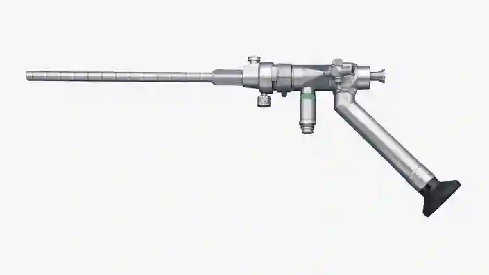
Visualization and Instruments for Endonasal Skull Base Surgery
- À la carte concept for efficient compilation of a customized instrument set
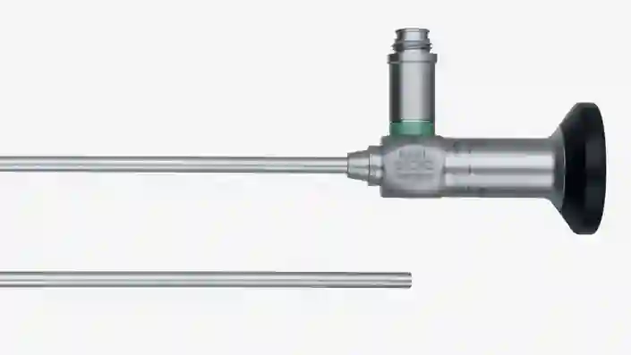
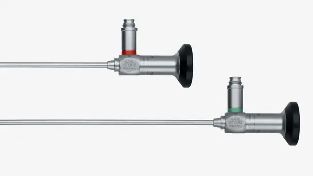
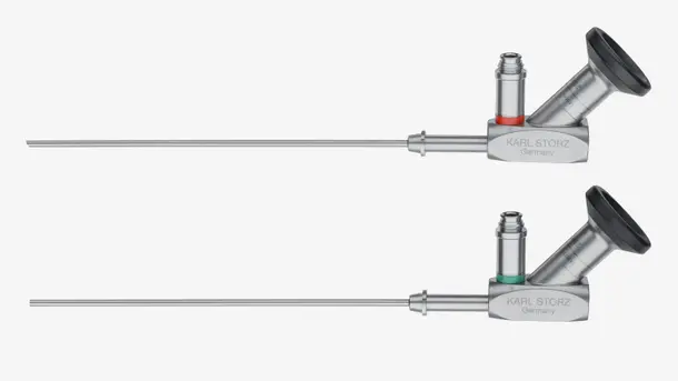
- Diverse 2D telescopes with different directions of view
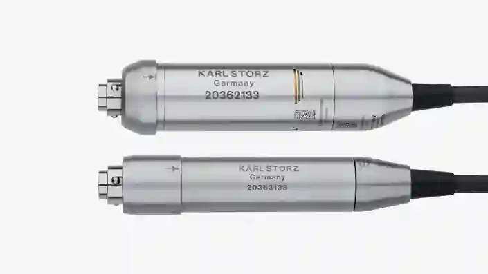
- TIPCAM®1 S 3D NEURO – 3D endoscopy in 0° and 30°
- Continuous vision thanks to effective lens cleaning systems
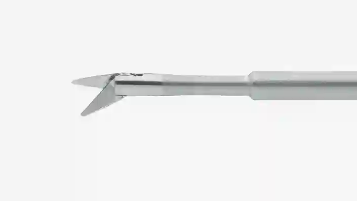
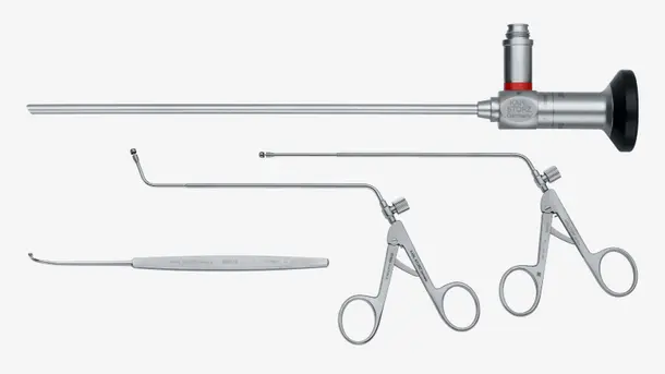
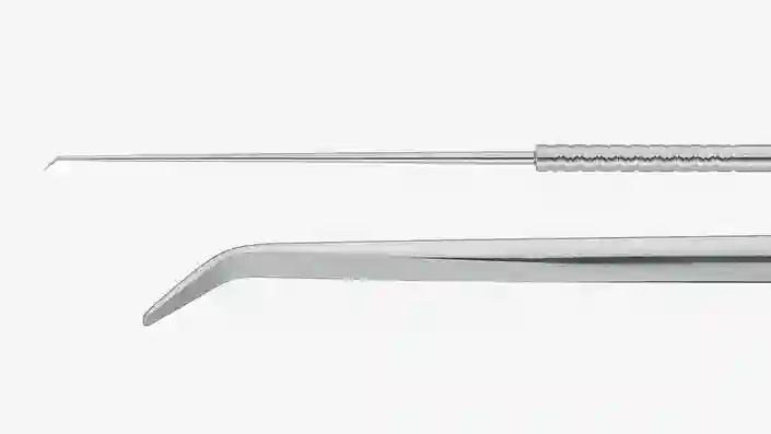
- Wide range of standard instruments such as curettes, dissectors, scissors, hooks and knives
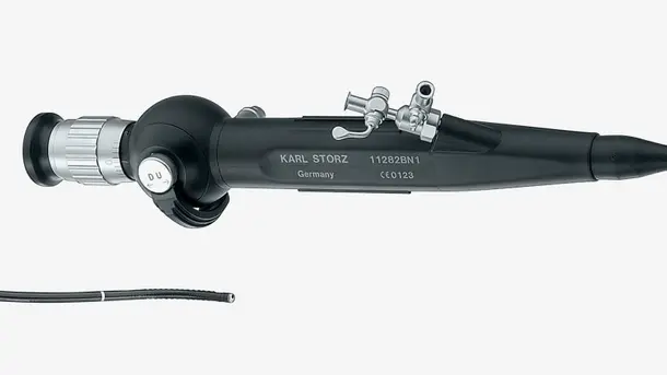
Multimodal Visualization – NIR/ICG Vascular Fluorescence-assisted Endoscopy in Neurosurgery
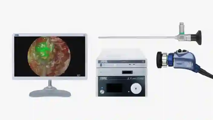
NIR/ICG fluorescence imaging – an OPAL1® technology from KARL STORZ – enables the visualization of vascular structures in real time. The inflow of indocyanine green (ICG) can be made visible with light at wavelengths in the near infrared range.
The use of endoscopic NIR/ICG technology and the administration of indocyanine green (ICG) enables neurosurgeons to identify anatomic structures.
This allows the intraoperative perfusion assessment of tissue, the identification of neurovascular structures, the differentiation of tissue layers between tumors and adjacent structures as well as the visual evaluation of blood vessels and their perfusion.1
The 4 mm NIR/ICG neuroendoscopes are available with diverse directions of view (0°, 30° and 45°) and can be used with both near infrared imaging and standard white light.
1 Catapano et al: Multimodal use of indocyanine green endoscopy in neurosurgery: a single-center experience and review of the literature, Neurosurgery Review, 2017
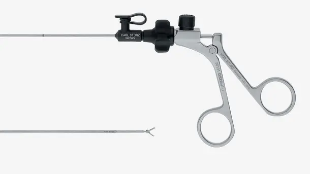
Visualization and Instruments for Keyhole Neurosurgery
- À la carte concept for efficient compilation of a customized instrument set
- VITOM® 3D system – a revolutionary solution for the visualization of microsurgical and open surgical interventions
- Angled 2D telescopes with thin diameters for space-saving keyhole neurosurgery
- TIPCAM®1 S 3D NEURO – 3D endoscopy in 0° and 30°
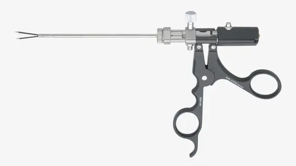
- Delicate and ergonomic single-sheath micro instruments with straight or bayonet-shaped design
- Unique TAKE-APART® bipolar forceps with various inserts
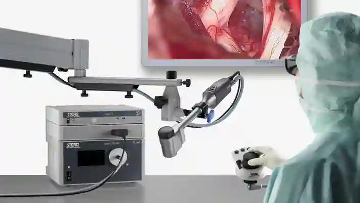
VITOM® 3D – 3D Visualization for Neurosurgery
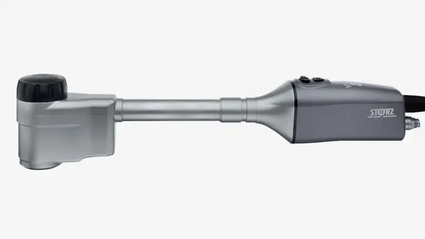
The VITOM® 3D system provides a solution for the visualization of microsurgical and open surgical interventions. Application possibilities are similar to that of the operating microscope.1 The system is controlled via the IMAGE1 PILOT which is mounted on the OR table in close proximity to the surgeon.
- Smaller, lighter and more compact than an operating microscope2
- Lower acquisition costs and creates synergistic effects with endoscopy by using the same video tower – thus combining the benefits of endoscopy and microscopy1
- Ergonomic work – the user is not confined to the eyepiece1
- Improved workflow – the OR team can view the procedure in the same image quality as the surgeon1
1 Oertel: Vitom-3D for Exoscopic Neurosurgery: Initial Experience in Cranial and Spinal Procedures,
World Neurosurgery (105), September 2017, p. 161
2 Siller: A high-definition 3-dimensional exoscope as an alternative to the operating microscope in spinal microsurgery
LOTTA® 6° for Intracranial Neurosurgery
Special Features:
- Ventriculoscope with a sheath diameter of 6.8 mm – for the entire range of endoscopic intracranial procedures1
- 6° angle of view
- The working channel (diameter 2.9 mm) and the side channels (diameter 1.6 mm) allow bimanual dissection
- A wide range of support features such as, for example, possibility of rotation in the sheath and instruments with length markings
1 Schroeder: Application of the LOTTA Ventriculoscopic System in Clinical Practice, 2014, p. 8
LOTTA® 30° for Intracranial Neurosurgery
Special Features:
- Ventriculoscope with 30° direction of view
- Sheath diameter 6.8 mm
- The working channel (diameter 2.9 mm) and side channels (diameter 1.6 mm) allow bimanual dissection
- A wide range of support features such as, for example, possibility of rotation in the sheath and instruments with length markings
LITTLE LOTTA® 6° for Intracranial Neurosurgery
Special Features:
- Ventriculoscope with a sheath diameter of 4.5 mm – particularly suitable for patients with small ventricles or narrow foramen of Monro as well as for tumor biopsies1
- 6° angle of view
- A wide range of support features such as, for example, possibility of rotation in the sheath and special instruments
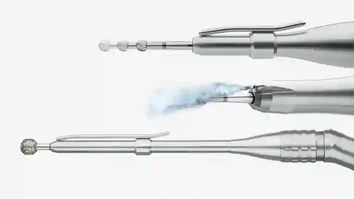
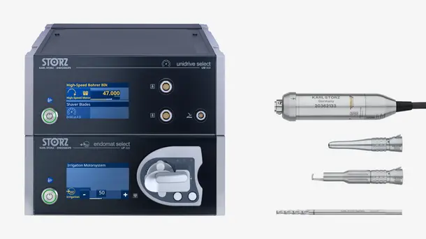
UNIDRIVE® S III* NEURO – High-Speed Motor System for Neuro and Spine Surgery
Special Features:
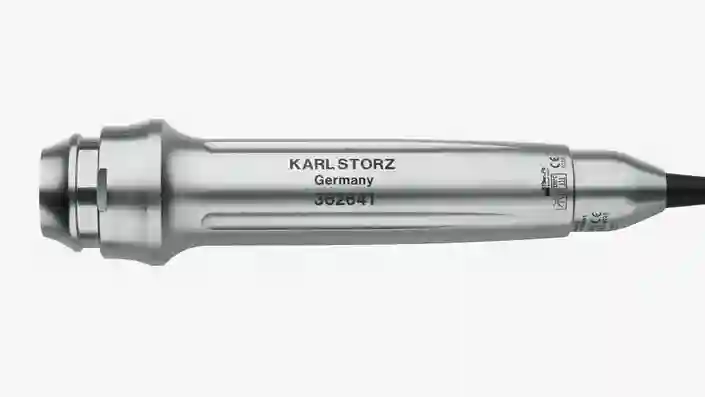
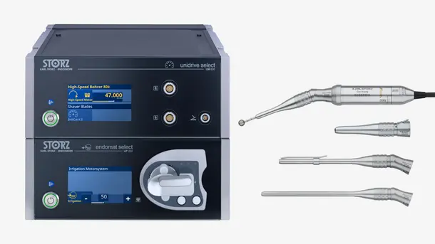
- High-speed handpieces with up to 100,000 rpm
- Selection of handpieces and shavers
- Integrated touch screen for menu guidance and program selection
- For opening the skull and subsequent craniotomy according to individual preferences by means of various dura protector or perforator attachments for the craniotomy handpiece
* not for sale in the U.S.