Neurosurgery
KARL STORZ has come a long way since being a pioneer in neuroendoscopic products during the 1990s. Today, we offer solutions to address intraventricular, cranial, and skull base pathologies. Together with our customers, we can envision better patient outcomes through exceptional imaging, instrumentation, and systems integration.
KARL STORZ has come a long way since being a pioneer in neuroendoscopic products during the 1990s. Today, we offer solutions to address intraventricular, cranial, and skull base pathologies. Together with our customers, we can envision better patient outcomes through exceptional imaging, instrumentation, and systems integration.
.svg)
.svg)

Solutions in the Spotlight
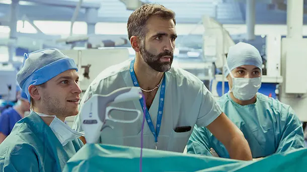
Healthcare professionals
We provide reliable, tailored and innovative solutions to overcome your surgical challenges and improve your clinical results in neurosurgery and spine surgery.

Finance & facilities
Our solutions help to expand neurosurgical treatments by attracting more patients with a greater range of services.

Patient outcome
Minimally invasive treatments have reported faster recovery rates and lower complication rate among patients.15
Highlights by Procedure
Endoscopic Skull Base Surgery
Everything you’ll need to get started
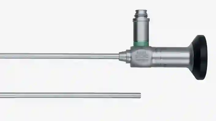
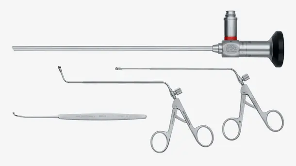
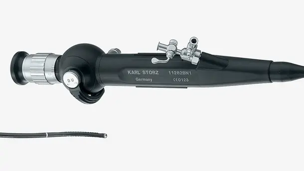
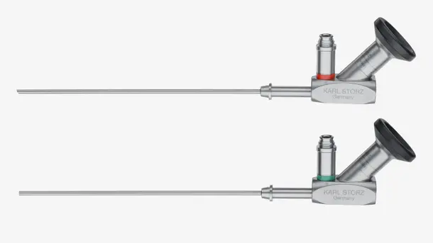
Skull base endoscopes
From 0°, 30°, 45° to 70°, straight and angled eyepieces with various diameters, 2D and 4K capabilities.
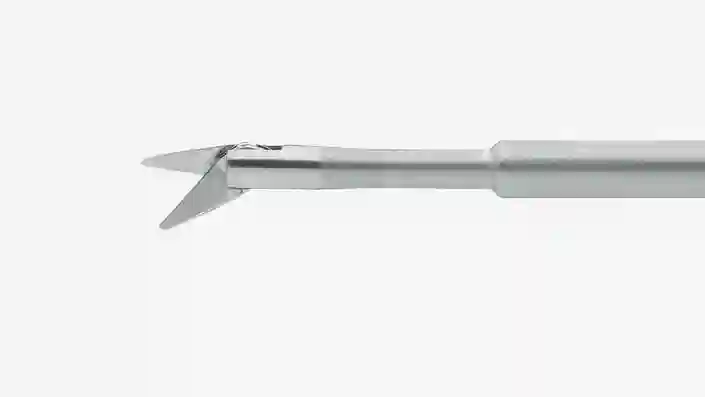
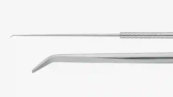
Skull base instruments
Explore our comprehensive set of specialized endoscopic skull base instrumentation.
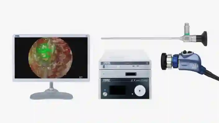
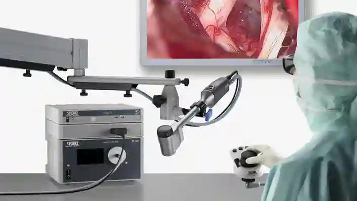
NIR/ICG in neurosurgery
The 4 mm endoscopic near-infrared indocyanine green of IMAGE1 S™ RUBINA™ helps identify critical neurovascular structures.67
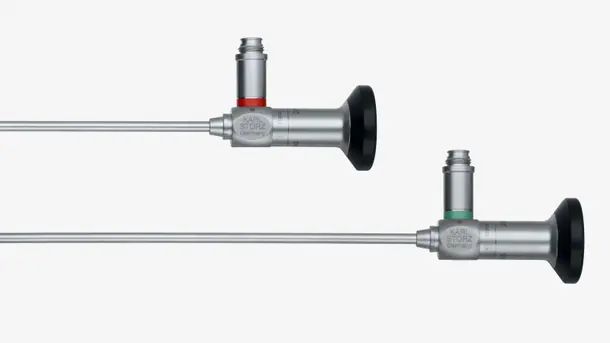
Highlights from our range
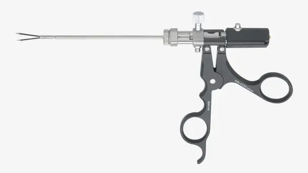
Intraventricular Neuroendoscopy
Our state-of-the-art intraventricular neuroendoscopy solutions for obstructive hydrocephalus and other lesions within the ventricular system are the result of our close partnership with experienced surgeons. By using our cutting-edge technology, you can avoid the need for shunts in your obstructive hydrocephalus patients1, resulting in improved quality of life.8
Everything you’ll need to get started
Highlights from our range

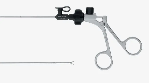
Endoscope-assisted & Keyhole Neurosurgery
Everything you’ll need to get started
Highlights from our range

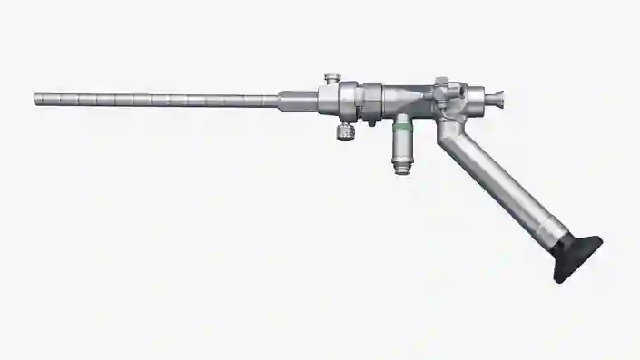
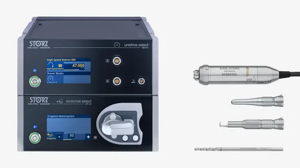
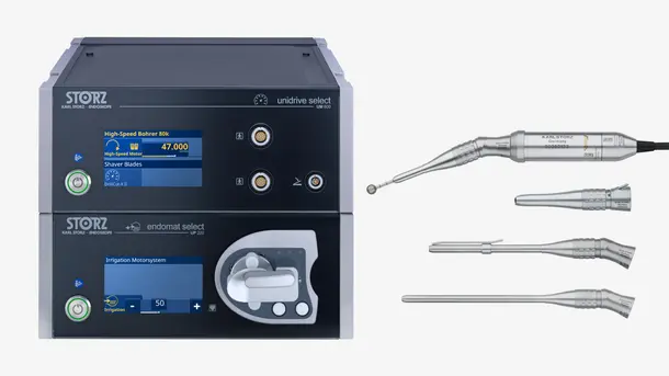
Surgical Motor System for Neurosurgery
Our UNIDRIVE™ SELECT multidisciplinary and modular motor system is designed to offer a range of customization possibilities along with synergistic benefits in cross-specialty integration.
Everything you’ll need to get started
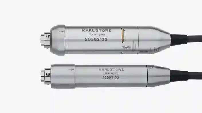
High-speed motors
Air-cooled HighDrive™ is designed to deliver consistent performance and comfort during long procedures. Compact, low-weight LightDrive is quiet even at high speeds.
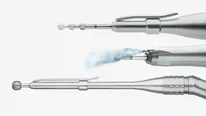
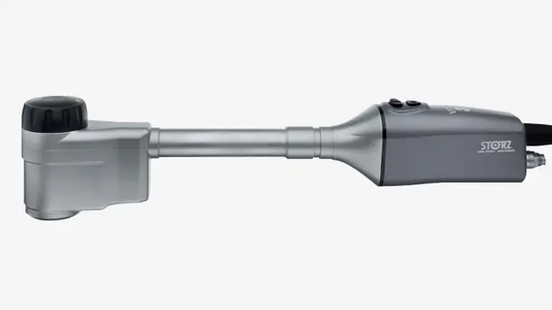
High-speed handpieces
Our high-speed handpieces come in many lengths and are compatible with HighDrive™ and LightDrive micro motors, with speeds up to 80,000 rpm.
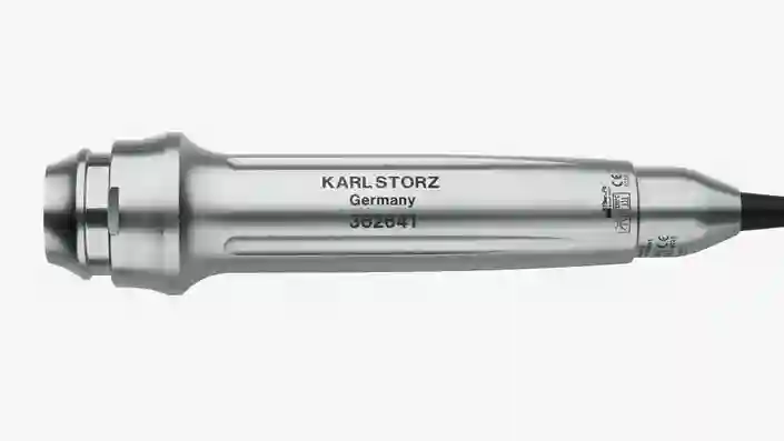
Perforator
The ergonomic perforator is designed for power and control during trepanation procedures.
Highlights from our range
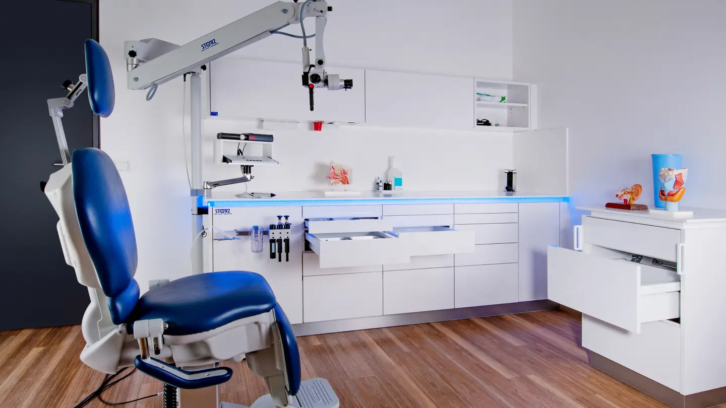
Create state-of-the-art surgical spaces tailored to your needs
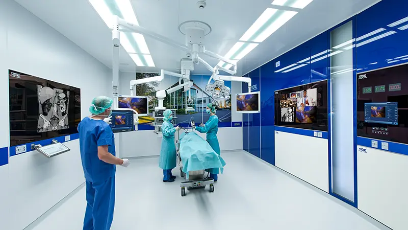
The future of operating room integration