
Les solutions d'imagerie
Fabricant de matériel et de systèmes endoscopiques, KARL STORZ propose des solutions modulaires et adaptables dont le but est de mettre à disposition de chaque client la chaîne d'image correspondant à ses besoins spécifiques.
Fabricant de matériel et de systèmes endoscopiques, KARL STORZ propose des solutions modulaires et adaptables dont le but est de mettre à disposition de chaque client la chaîne d'image correspondant à ses besoins spécifiques.
CARACTÉRISTIQUES
Précision - Modularité - Adaptabilité
.svg)


Produits phare


Les solutions d’imagerie
Nous mettons nos technologies d'imagerie au service de la visualisation aussi bien en salle d'opération qu'en cabinet médical. Paramétrable, la chaîne d'image s'adapte aux besoins individuels de chacun de nos clients.
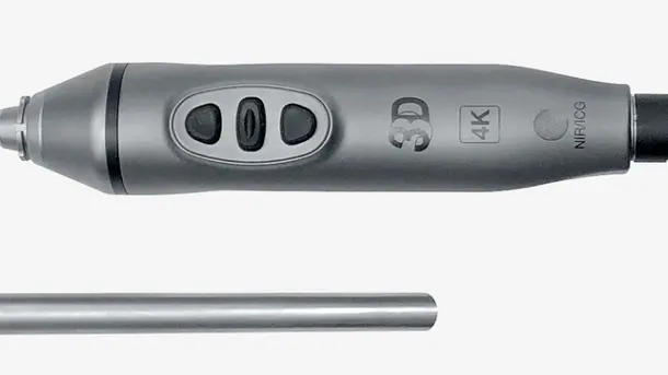
IMAGE1 S™ Rubina®
L'alliance des technologies 4K et 3D à l'imagerie par fluorescence NIR/ICG contribue à renforcer la fiabilité de détection et la précision chirurgicale. Le principe NIR/ICG permet d'élargir l'éventail des possibilités comme, par exemple, évaluer la circulation sanguine ou encore déceler les ganglions sentinelles.
Les modes de visualisation
Grâce aux différents modes de visualisation, les structures anatomiques sont nettement différenciables les unes des autres.
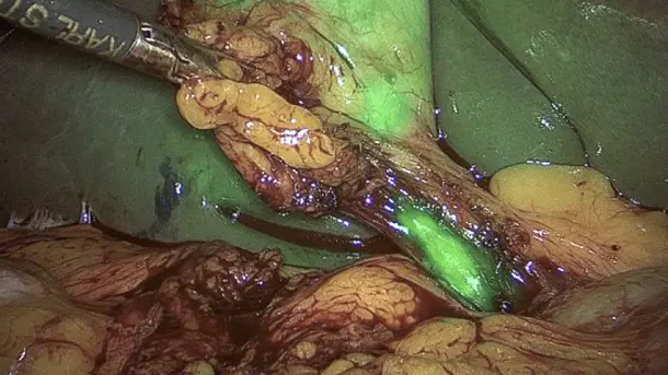
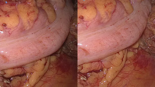
Mode incrustation « Overlay »
En mode incrustation, l'image caméra classique en spectre visible est combinée avec les informations NIR/ICG afin de générer une image superposée.
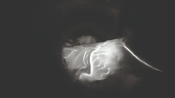
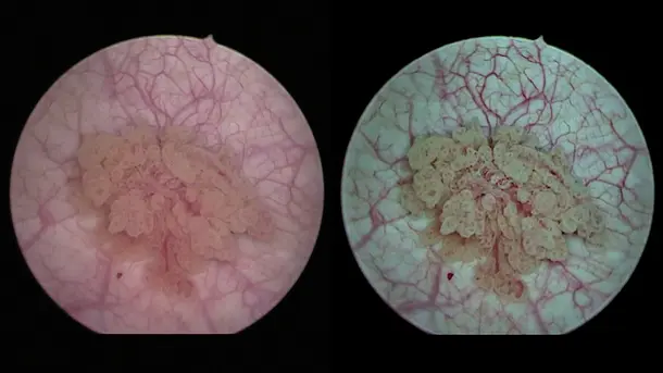
Représentation monochromatique
Le signal NIR/ICG s'affiche en blanc sur fond noir afin de mettre en relief les marges structurelles.
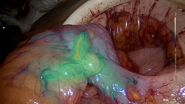
Affichage intensité du signal « Intensity Map »
Affichage de l’intensité du signal NIR/ICG au moyen d’un gradient colorimétrique
CLARA
Cette Technologie S fournit un éclairage homogène, empêche les phénomènes de surexposition et de réflexion tout en favorisant la visibilité dans les zones sombres.
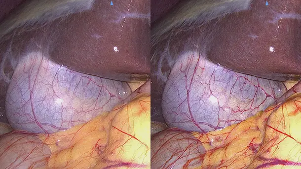
CHROMA
Cette Technologie S accentue le contraste des couleurs sans en modifier la perception naturelle. Les changements de couleur et les structures sont mis en relief.
CLARA + CHROMA
Utilisées en combinaison, ces Technologies S fournissent une image uniformément lumineuse et mettent en relief les structures tissulaires.
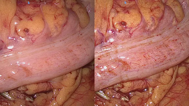
SPECTRA A
Cette Technologie S permet le filtrage spectral des tons rouges. Les structures rouges les plus fines, comme les vaisseaux sanguins et les muqueuses, sont représentées de façon marquée dans une teinte vert-bleue.
SPECTRA B
Cette Technologie S contribue à réduire les tons rouges tout en amplifiant la partie vert-bleue du spectre chromatique. L’arrière-plan de l’image apparaît dans une couleur verdâtre faisant ressortir les vaisseaux et les capillaires sanguins.
Les produits de la gamme IMAGE1 S™ Rubina®
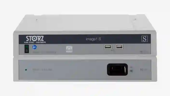
IMAGE 1 S™
Nos solutions de visualisation fournissent l'expérience visuelle nécessaire à une prise en charge opératoire optimale de chaque patient. Conçus selon les principes de modularité et de durabilité, nos systèmes de visualisation sont adaptables aux besoins individuels.
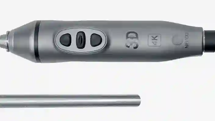
TIPCAM®1 Rubina®
Ce vidéo-endoscope « All-in-one » (tout-en-un) combine les 3 technologies d'imagerie 4K, 3D et NIR/ICG dans un seul produit pour fournir une visualisation de qualité et une perception naturelle des profondeurs.
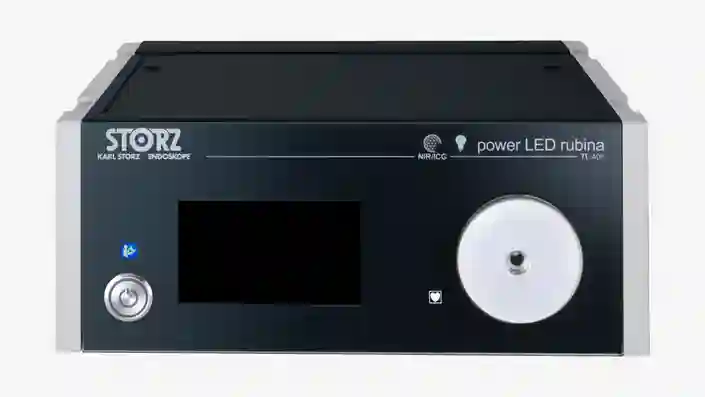
Power LED Rubina®
Cette source de lumière froide sans laser a été conçue pour les applications NIR/ICG et dans le spectre visible.
Un système d'imagerie NIR/ICG évolutif
Des systèmes adaptés aux besoins spécifiques grâce au principe de compatibilité des composantes entre elles
Les domaines d’application IMAGE1 S™ Rubina®
"Le système IMAGE1 S™ Rubina® nous a permis de faire un grand pas en avant dans le domaine de la chirurgie assistée par fluorescence. Nous sommes comme « sortis de l'obscurité » pour entrer dans la lumière 3D 4K, ce qui nous permet de traiter les patients dans de meilleures conditions."

IMAGE1 S™ 4U
Pour KARL STORZ, la qualité d'image est synonyme d'excellence. Dans cet esprit, la définition 4K a logiquement été associée à la visualisation endoscopique.
Les modes de visualisation
Différents modes de visualisation permettent d'identifier clairement et de différencier nettement les structures anatomiques.
CLARA
Cette Technologie S fournit un éclairage homogène, empêche les phénomènes de surexposition et de réflexion tout en favorisant la visibilité dans les zones sombres.
CHROMA
Cette Technologie S accentue le contraste des couleurs sans en modifier la perception naturelle. Les changements de couleur et les structures sont mis en relief.
CLARA + CHROMA
Utilisées en combinaison, ces Technologies S fournissent une image uniformément lumineuse et mettent en relief les structures tissulaires.
SPECTRA A
Cette Technologie S permet le filtrage spectral des tons rouges. Les structures rouges les plus fines, comme les vaisseaux sanguins et les muqueuses, sont représentées de façon marquée dans une teinte vert-bleue.
SPECTRA B
Cette Technologie S contribue à réduire les tons rouges tout en amplifiant la partie vert-bleue du spectre chromatique. L’arrière-plan de l’image apparaît dans une couleur verdâtre faisant ressortir les vaisseaux et les capillaires sanguins.
Les produits de la gamme IMAGE1 S™ 4U
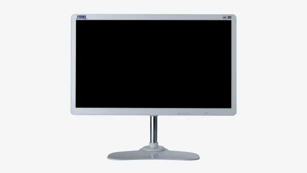
IMAGE 1 S™
Nos solutions de visualisation fournissent l'expérience visuelle nécessaire à une prise en charge opératoire optimale de chaque patient. Conçus selon les principes de modularité et de durabilité, nos systèmes de visualisation sont adaptables aux besoins individuels.
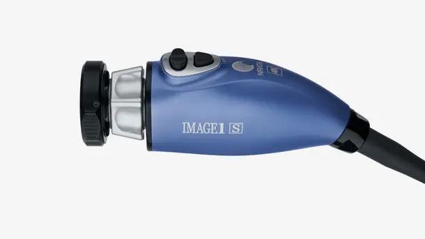
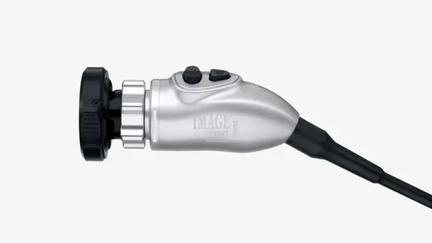
La tête de caméra IMAGE1 S™ 4U
Cette tête de caméra offre le format 4K ainsi qu'un espace colorimétrique étendu afin de pouvoir identifier clairement et différencier nettement les structures anatomiques les unes des autres.
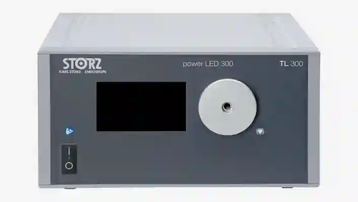
POWER LED 300
Cette source de lumière froide sans laser a été conçue pour les applications dans le spectre visible.
Un système d'imagerie 4K évolutif
Des systèmes adaptés aux besoins spécifiques grâce au principe de compatibilité des composantes entre elles
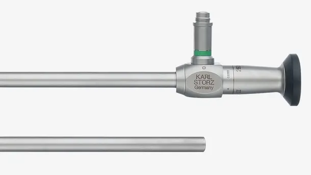
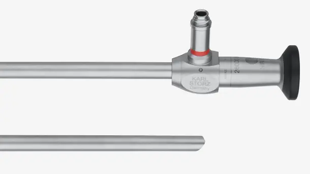
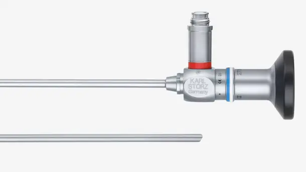
Les optiques HOPKINS®
Nos optiques sont conçues pour les applications dans le spectre visible.
Les domaines d’application IMAGE1 S™ 4U
"La technologie IMAGE1 S™ nous a soudainement permis d'apercevoir des choses qu'il ne nous avait pas été donné de voir auparavant. Une visualisation de qualité contribue à améliorer les possibilités de traitement."

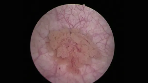
IMAGE1 S™ Saphira™
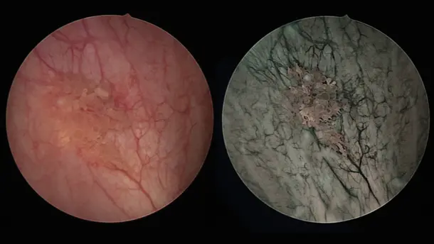
Grâce au procédé Blue Light Imaging BLI - Imagerie lumière bleue - (anciennement PDD) utilisé suite à l'administration de la substance Hexvix/Cysview, les tumeurs malignes à un stade précoce sont visualisables, à la différence du diagnostic sous lumière blanche qui ne permet pas, ou difficilement, de les détecter.
Les modes de visualisation
Différents modes de visualisation contribuent à différencier nettement les structures anatomiques.
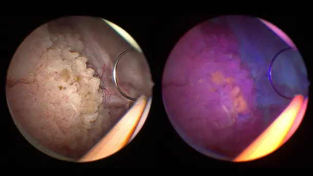
BLI
La lumière bleue permet de distinguer plus aisément les cellules tumorales (en rouge fluorescent) par rapport aux tissus environnants (en bleu).
CHROMA
Cette Technologie S accentue le contraste des couleurs sans en modifier la perception naturelle. Les changements de couleur et les structures sont mis en relief.
SPECTRA A
Cette Technologie S permet le filtrage spectral des tons rouges. Les structures rouges les plus fines, comme les vaisseaux sanguins et les muqueuses, sont représentées de façon marquée dans une teinte vert-bleue.
SPECTRA B
Cette Technologie S contribue à réduire les tons rouges tout en amplifiant la partie vert-bleue du spectre chromatique. L’arrière-plan de l’image apparaît dans une couleur verdâtre faisant ressortir les vaisseaux et les capillaires sanguins.
Les produits de la gamme IMAGE1 S™ Saphira™
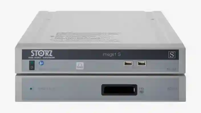
IMAGE 1 S™
Nos solutions de visualisation fournissent l'expérience visuelle nécessaire à une prise en charge opératoire optimale de chaque patient. Conçus selon les principes de modularité et de durabilité, nos systèmes de visualisation sont adaptables aux besoins individuels.
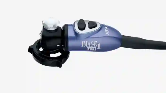
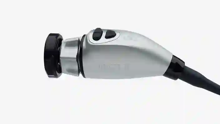
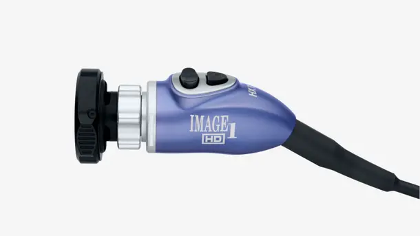
La tête de caméra IMAGE1 S™ HX-P FI
Légère et ergonomique, cette tête de caméra pendulaire a été conçue pour les applications Blue Light Imaging (lumière bleue) et dans le spectre visible.

POWER LED Saphira™
Cette source de lumière froide sans laser a été conçue pour les applications dans le spectre visible et l'imagerie Blue Light (sous lumière bleue).
L'imagerie Blue Light : une solution évolutive
Des systèmes adaptés aux besoins spécifiques grâce au principe de compatibilité des composantes entre elles
Les optiques HOPKINS® BLI
Nos optiques compatibles sont destinées aux applications Blue Light (sous lumière bleue) et dans le spectre visible
Les domaines d’application IMAGE1 S™ Saphira™
« J'ai eu l'opportunité d'utiliser la nouvelle source de lumière Saphira™ au cours des derniers mois et peux affirmer qu'elle contribue à améliorer énormément la qualité de la procédure. »

TELE PACK+
Conçue pour le diagnostic et les interventions mineures, cette plateforme compacte est indiquée pour l'usage en cabinet médical, en clinique de jour ainsi qu'au service des urgences, au service des soins intensifs ou encore pour le traitement en ambulatoire.
La gamme des produits TELE PACK+
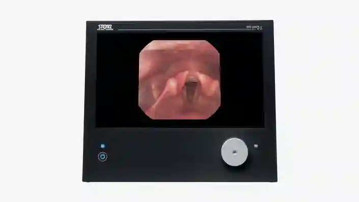
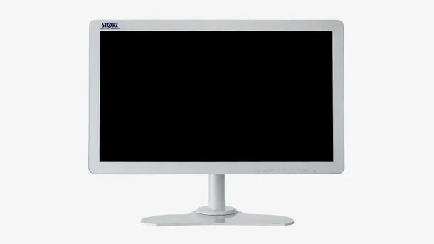
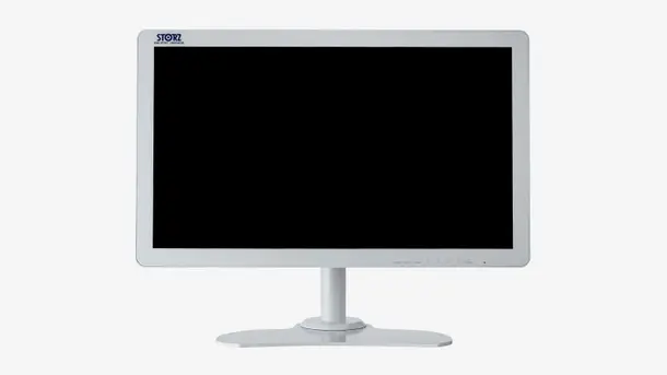
TELE PACK+
Ce système réunit un écran, une source de lumière LED, une unité de commande caméra et une fonction de documentation avec option réseau dans un appareil compact et portable.

X-Line et C-Line
Grâce à ses ports X-Line et C-Line, le TELE PACK+ s'utilise avec différents modèles d'endoscope rigide, flexible et à usage unique dans de nombreuses disciplines médicales.
Un système d'imagerie FULL HD compact évolutif
Des systèmes adaptés aux besoins spécifiques grâce au principe de compatibilité des composantes entre elles
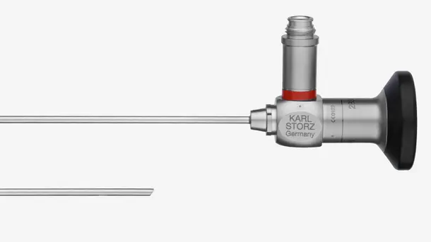
Les endoscopes rigides
Nos optiques rigides sont conçues pour les applications dans le spectre visible.
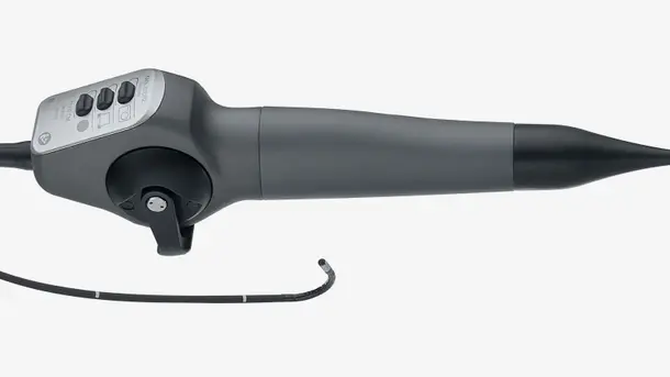
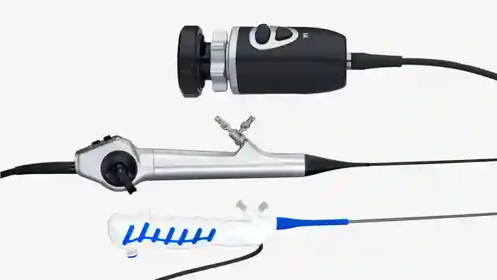
Les vidéo-endoscopes
Nos vidéo-endoscopes flexibles permettent de visualiser les cavités les plus étroites et les plus sinueuses sans avoir recours à aucun procédé invasif. Notre gamme de vidéo-endoscopes propose des modèles pour quasiment toutes les disciplines médicales.
Les domaines d’application de TELE PACK+
"Nous apprécions énormément le nouveau système TELE PACK+ car la qualité des images qu'il fournit permet de livrer un diagnostic efficace et fiable même dans les cas difficiles."

TELECAM C3
L'unité de commande caméra TELECAM C3 s'utilise pour le diagnostic et les interventions mineures en cabinet médical ou dans l'environnement chirurgical. Conçue pour l'emploi avec différents types d'endoscope, elle est utilisable dans de nombreuses spécialités médicales.
Les produits TELECAM C3
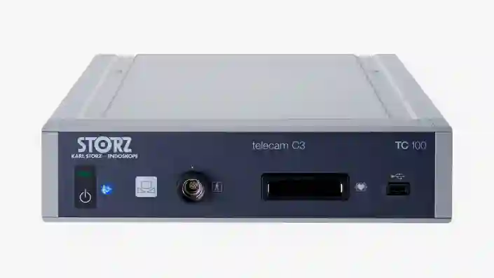
TELECAM C3
Destinée aux interventions endoscopiques simples, cette unité de commande caméra compacte est équipée de deux raccords caméra qui lui permettent d'être utilisée conjointement avec différents endoscopes.
X-Line et C-Line
Grâce à ses ports X-Line et C-Line, la TELECAM C3 s'utilise avec différents modèles d'endoscope rigide, flexible et à usage unique dans de nombreuses disciplines médicales.
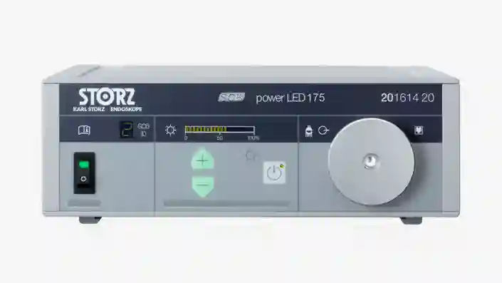
POWER LED 175
Utilisable pour illuminer les petites cavités, cette source de lumière froide sans laser a été conçue pour les applications dans le spectre visible.
Un système d'imagerie FULL HD évolutif
Des systèmes adaptés aux besoins spécifiques grâce au principe de compatibilité des composantes entre elles
Les endoscopes rigides
Nos optiques rigides sont conçues pour les applications dans le spectre visible.
Les vidéo-endoscopes
Nos vidéo-endoscopes flexibles permettent de visualiser les cavités les plus étroites et les plus sinueuses sans avoir recours à aucun procédé invasif. Notre gamme de vidéo-endoscopes propose des modèles pour quasiment toutes les disciplines médicales.
Les domaines d’application de la TELECAM C3
Un environnement opératoire élaboré sur-mesure pour répondre aux besoins individuels
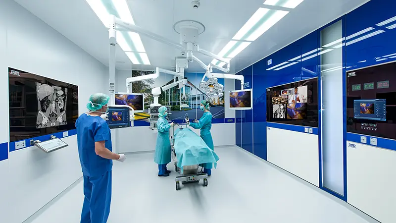
La salle d'opération intégrée
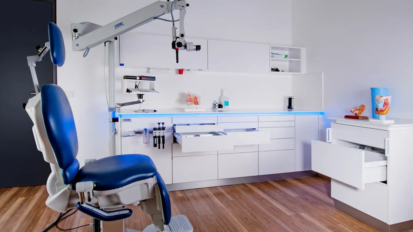
Le principe modulaire en cabinet médical
Savoir c'est pouvoir
La mise au point de techniques chirurgicales de plus en plus complexes ainsi que la conception d'instruments et d'appareils ultra-sophistiqués rendent nécessaires les besoins en formation continue tout en ouvrant de nouvelles perspectives d'évolution professionnelle. Nous mettons à la disposition des médecins et assistants médicaux dans le monde entier des cours et des ateliers de formation.


CONTACT Prendre contact avec nos spécialistes
Nos spécialistes vous conseillent dans le choix des produits répondant à vos attentes et vos besoins.