Secondary Prevention of Cervical Cancer
Intervascular Distance – Cancer
Atypical vessels separated by neoplastic epithelium indicate cancer.
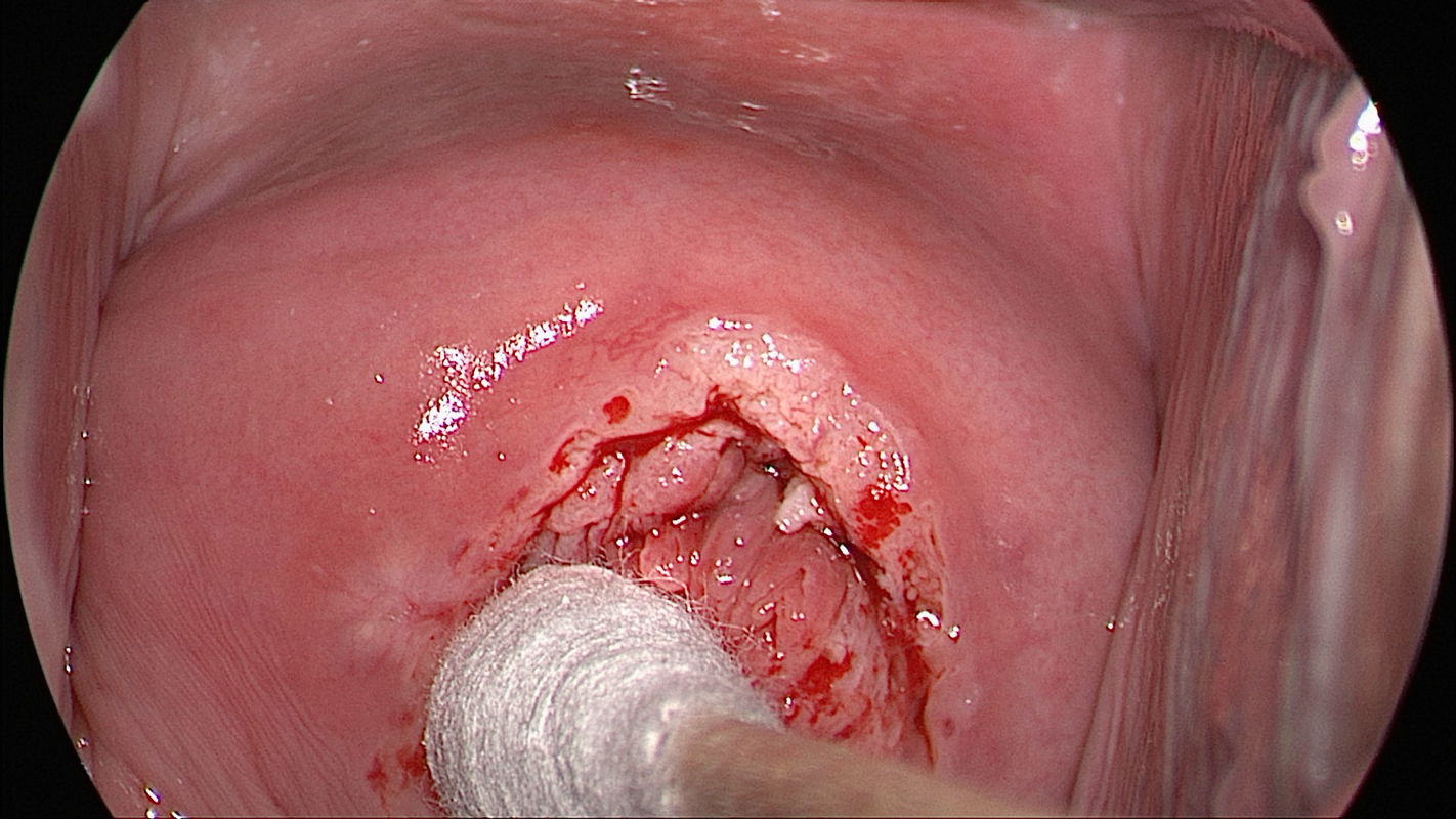
A high degree of cell proliferation and neovascularity may cause atypical vessels to be spaced farther apart, suggesting the presence of invasive cancer. Colposcopic findings in a 24-year-old gravida 0 are presented. Her cervical smear was reported as class V-g, and HPV 18 was detected. Colposcopy shows an opaque, acetowhite atypical T-zone type 3, which is classified as abnormal colposcopic finding suspicious for invasive cancer. Atypical vessels with a large intervascular distance are visible at 12 o’clock through the intact squamous epithelium.
Tumor vessels are numerous and separated by stroma in invasive adenocarcinoma.
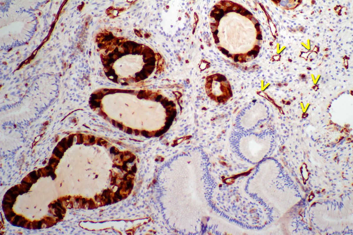
Histopathology of tissue sampled by loop biopsy shows highly atypical crypts (glands) whose epithelium expresses high levels of the marker stathmin 1 (STMN1). Normal endocervical crypts (glands) between 3 and 6 o’clock do not express STMN1. Within the stroma are numerous vessel lumina (arrowheads) whose endothelium is clearly marked by a positive STMN1 reaction. The histopathologic diagnosis is invasive endocervical adenocarcinoma G2. The patient was treated by laparoscopic sentinel lymphadenectomy and radical vaginal trachelectomy and was staged as pT1b1 pN0 (0/2 sn) G2 L0