Secondary Prevention of Cervical Cancer
Rag Sign
The advertisement is detached from the billboard.

Rags of tissue at the uterine cervix.
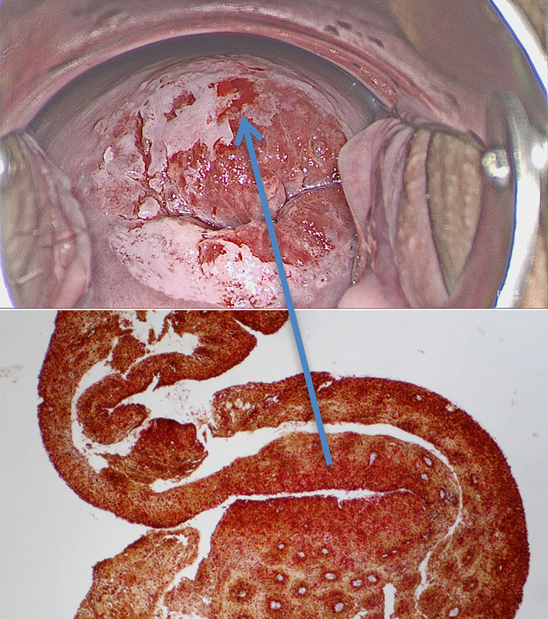
Rags of tissue are also seen at the uterine cervix when epithelium comes off in strips: rag sign is verified on the colpophotogram and the rag of tissue exhibits histologically all features of
HSIL detach easily from underlying connective tissue.
The rag sign is a fourth pathognomonic sign for high-grade SIL.
Colposcopic findings in a 35-year-old gravida 0 with no prior history of disease. The smear was reported as class III-p, and HPV 16 was detected. Colposcopy shows a circular acetowhite lesion with ‘ragged’ epithelium at 6 o’clock in an atypical T-zone type 2, classified as grade 2 (major change) abnormal colposcopic findings.
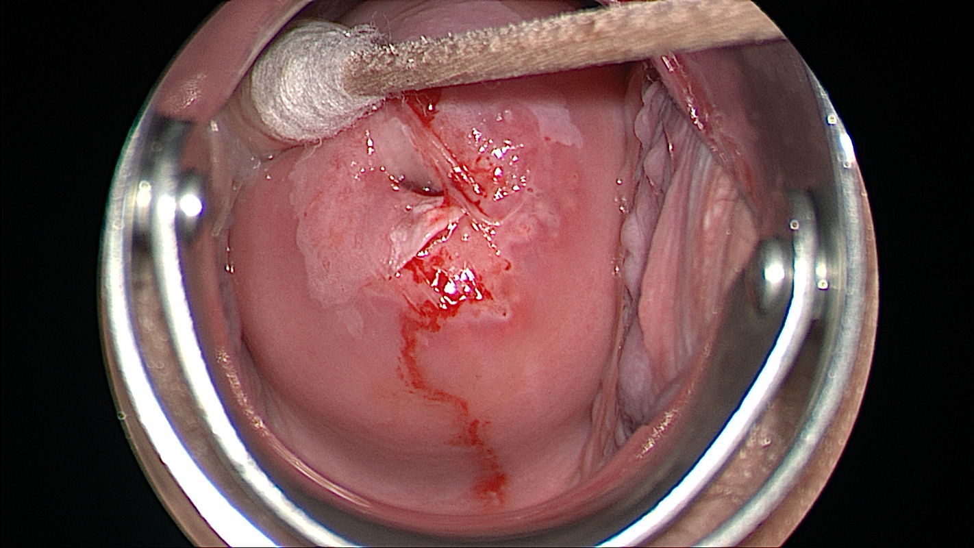
The detached strip of epithelium is sufficient for histological diagnosis.
The ragged epithelial strip was retrieved with a forceps. Histologic workup of the specimen shows atypical cells that occupy the full width of the epithelium and express high concentrations of the immune markers p16 and Ki67 into the highest cell layer.
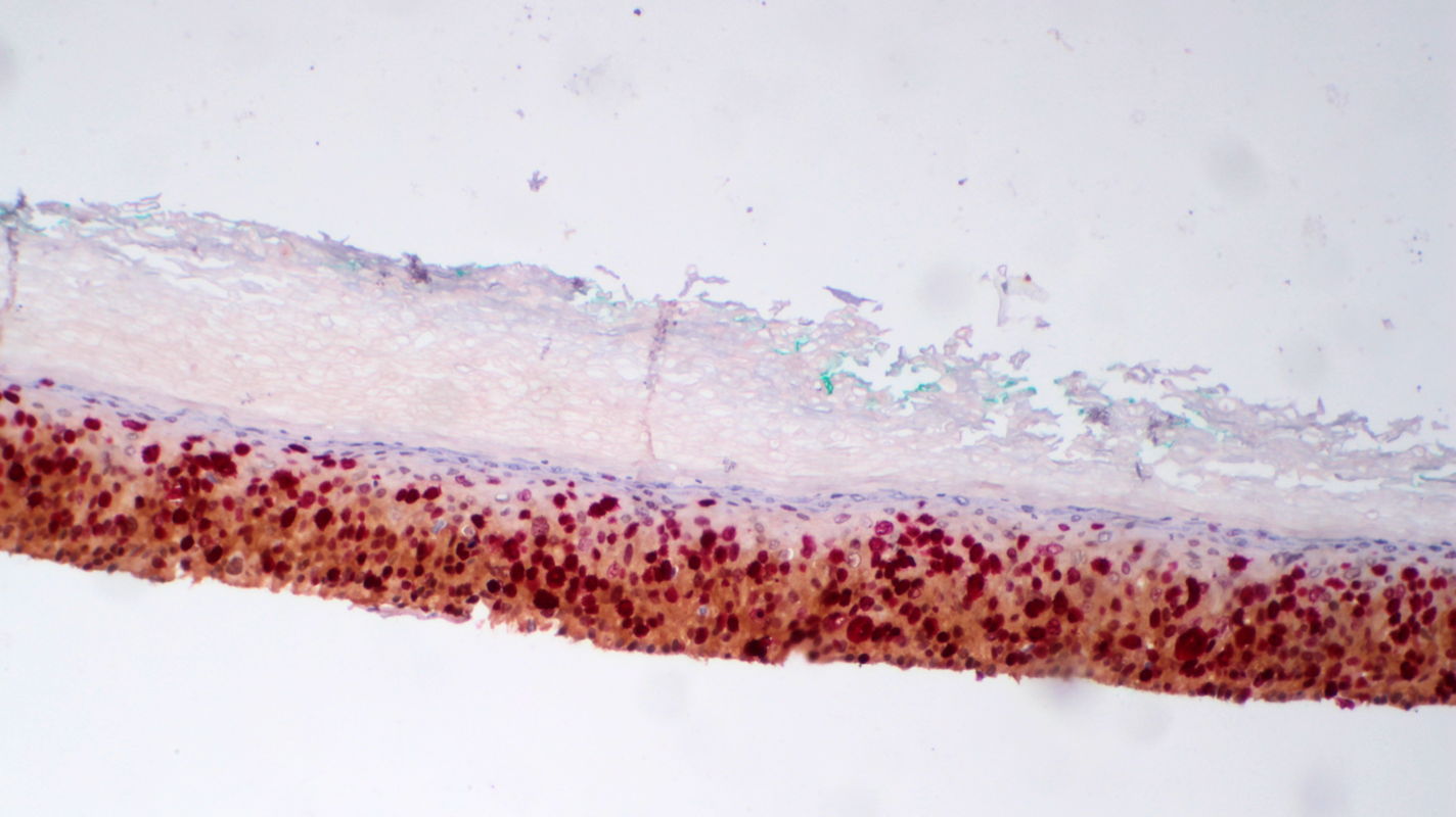
A non-nucleated keratin layer covers the epithelium superficially. The diagnosis is grade 3 cervical intraepithelial neoplasia (CIN 3).