Secondary Prevention of Cervical Cancer
Inner Border Sign

The ceiling light defines areas of varying brightness which result in several ‘borders’.
Borders between lesions are also seen on the uterine cervix: the border sign is identified following Lugol application and histologically a sharp border between metaplasia and
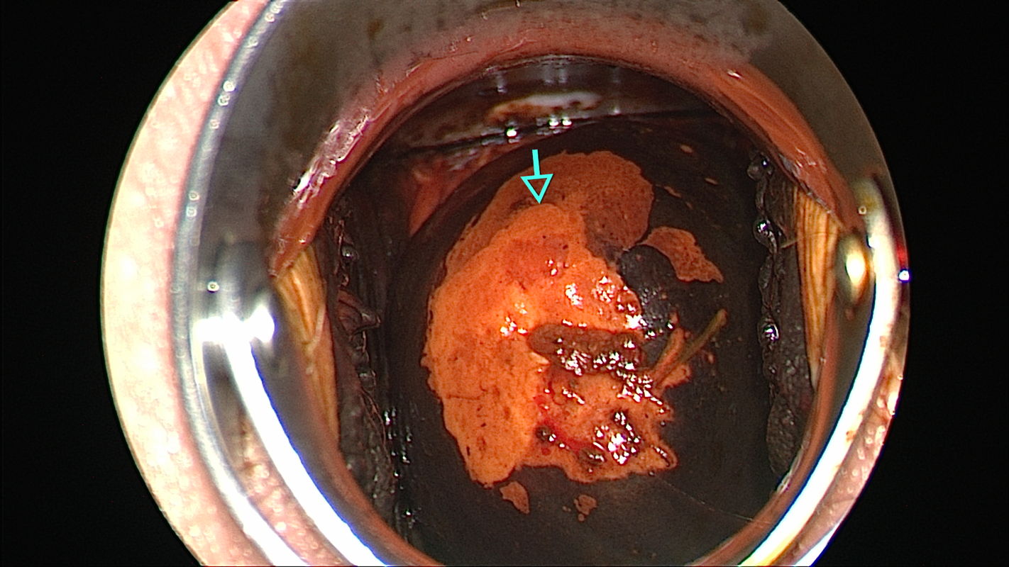
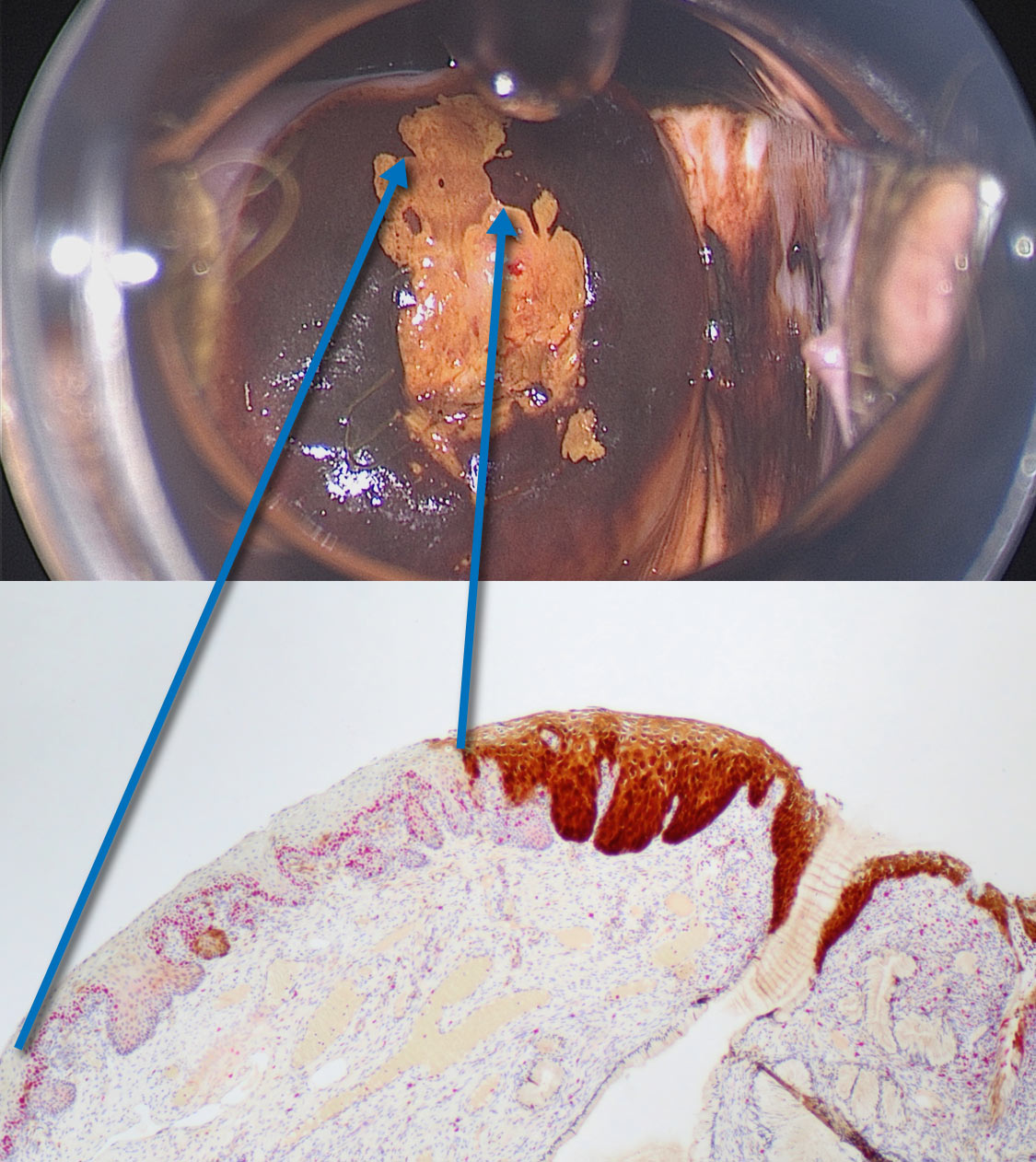
The inner border sign is considered pathognomonic for high-grade SIL. Colposcopic findings in a 35-year-old gravida 0 with no prior history of disease. The smear was reported as class III-p, and HPV 16 was detected. Colposcopy shows a circular lesion with partial iodine uptake at 11 to 1 o’clock and at 5 to 7 o’clock with a second, central lesion located between 6 and 2 o’clock in an atypical T-zone type 2. The central lesion is classified as grade 2 (major change) abnormal colposcopic finding.
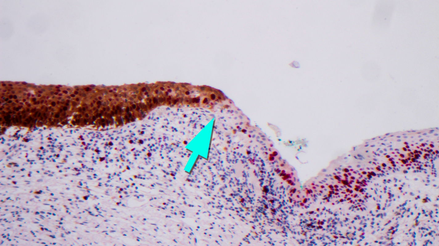
Tissue was sampled by loop excision in the transitional area between the two lesions. Histopathology of the central lesion (left of the arrow) shows atypical cells that uniformly occupy the full width of the epithelium and express high concentrations of the p16 and Ki67 immune markers into the highest cell layer. The diagnosis is grade 3 cervical intraepithelial neoplasia (CIN 3). This epithelium is abruptly replaced (right of the green arrow) by epithelium whose cells express Ki67 only into the middle epithelial layer and do not express p16. The diagnosis is grade 1 cervical intraepithelial neoplasia (CIN 1).