Secondary Prevention of Cervical Cancer
Adenocarcinoma
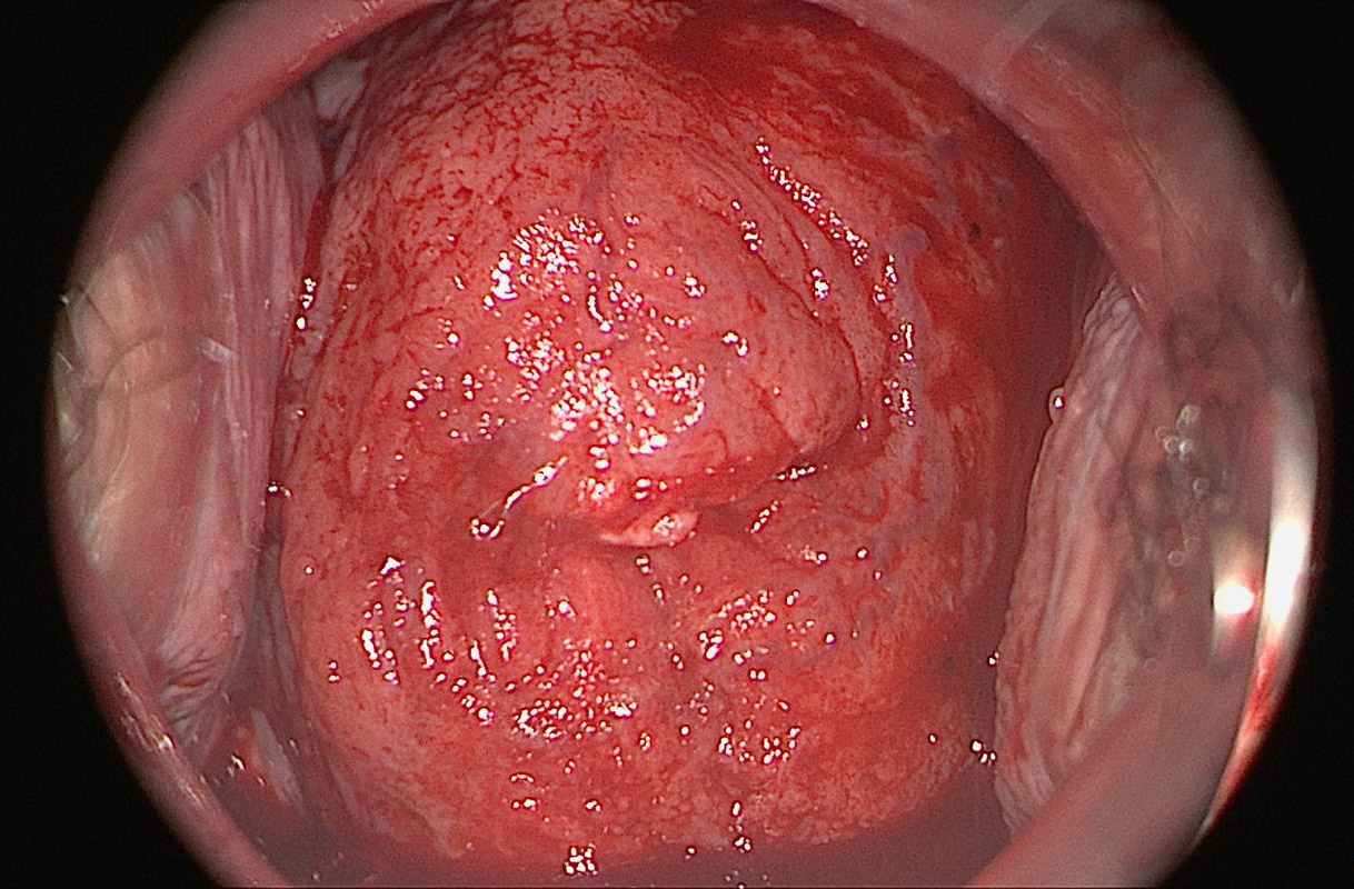
Atypical vessels associated with adenocarcinoma are indistinguishable from squamous cell carcinoma based on their external appearance.
Colposcopic findings in a 33-year-old gravida 0 with no prior history of disease. The smear was reported as class III-g, and HPV 18 was detected. Atypical vessels are found in an atypical T-zone type 1, classified as abnormal colposcopic finding suspicious for cancer.

Tissue sampled by punch biopsy shows atypical glandular complexes, which are classified as AIS between 11 and 6 o’clock and as invasive adenocarcinoma between 7 and 11 o’clock. All the glandular cells express high concentrations of the markers p16 and Ki67.
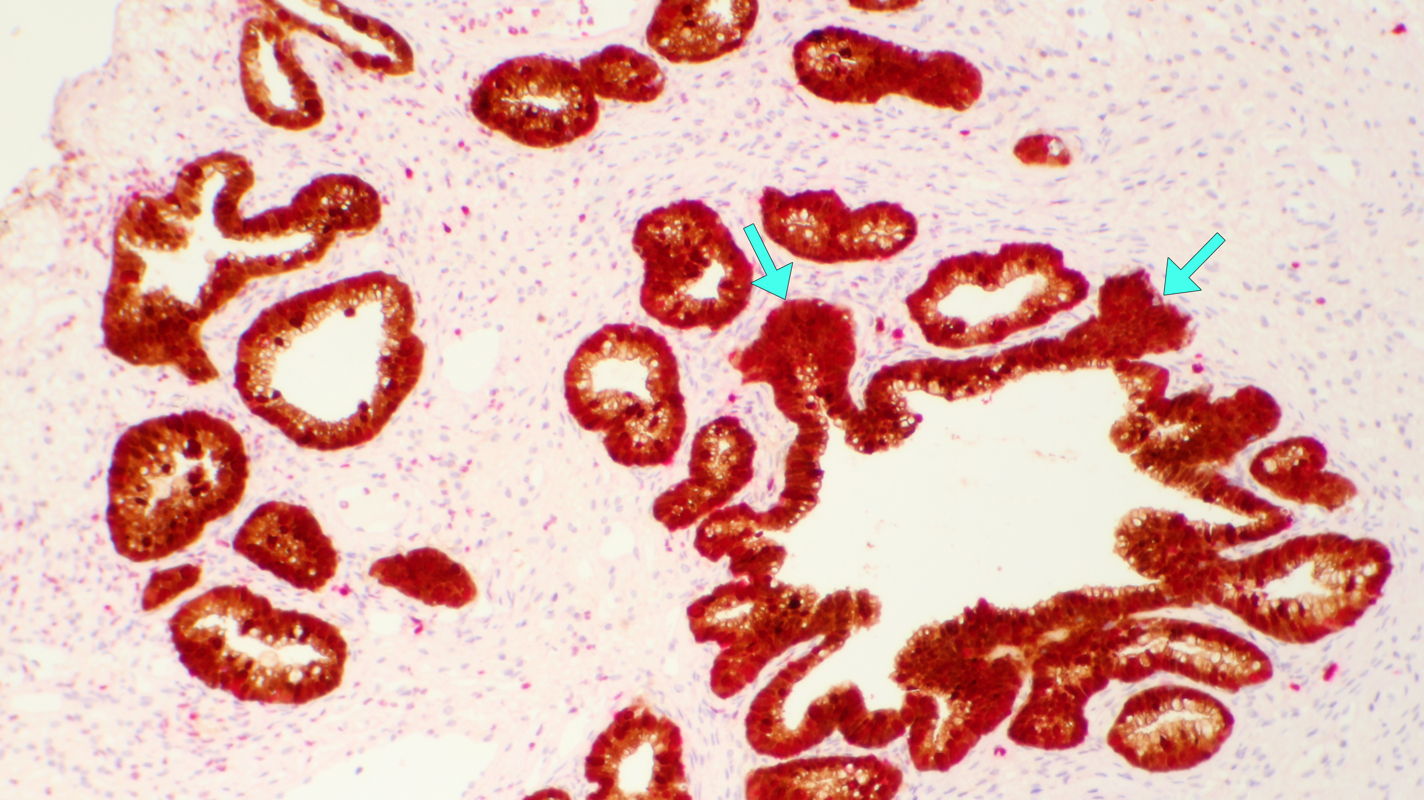
Magnified view of the invasive tumor component shows atypical cell complexes that have penetrated the basement membrane (green arrows). After a nerve-sparing laparoscopic radical hysterectomy, the disease was finally staged as pT1b2 pN0 (0/23) G2 L1 V0 Pn0.