Secondary Prevention of Cervical Cancer
Natural History of a Cervical HPV Infection
Topgraphy of HPV Attack of the Cervic
Natural History of a Cervical HPV Infection
Topgraphy of HPV Attack of the Cervic
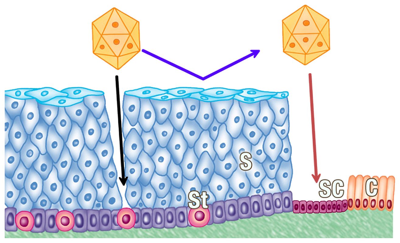
HPV and squamocolumnar junction cells are the origin of cervical neoplasia.
The schematic diagram shows the squamocolumnar junction junction, which is composed of non-keratinizing squamous epithelium (S) on the left and columnar epithelium (C) on the right. The cells in between are SC cells (SC in red) which are the main target of HPV on the cervix (red arrow). This is due to the SC cells lying exposed to the attacking HPVs on the outer surface. Unlike SC cells, the intact cell structure of squamous epithelium is capable of withstanding an HPV attack (blue arrow). In order to successfully accomplish an attack, a breach or lesion is needed that allows the virus to reach its target (black arrow) that are the rounded cells in the basal layer with stem cell (St) character. The gene expression profile of the discrete population of SC cells is different from that of neighbouring cells and shares common features with CIN and invasive cancers.