Secondary Prevention of Cervical Cancer
Histological Evaluation for CIN
Histological Evaluation for CIN
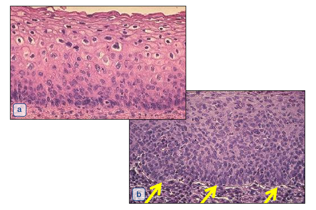
Precancerous lesions of the cervix are changes of the epithelium and not of the stroma.
Therefore, the severity of the precancerous lesions can be evaluated on the epithelium without stroma. The interaction between epithelium and stroma is only important in invasive disease. The histophotographs show CIN 1 (LSIL) (a) and CIN 3 (HSIL) (b). The histopathological appearance of individual cells of CIN 3 mimicks invasive cancer, however in CIN 3, the basal layer, marked by yellow arrows (b) is intact, which excludes invasive disease.
a) Differentiation in LSIL.
b) No differentiation in (HSIL).
CIN 3 and Atypical Mitotic Figures
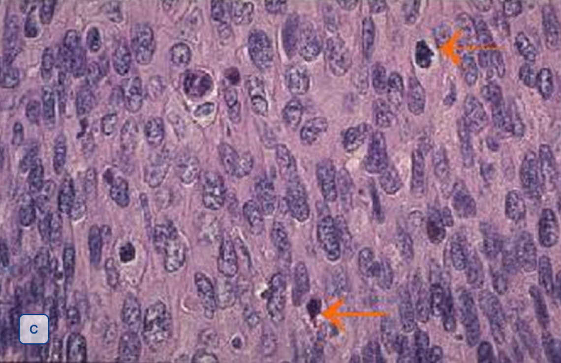
Besides the uniformity of atypical cells in all layers with increased nuclear-to-cytoplasmic ratio, the presence of atypical mitotic figures is the most important criterion for diagnosis of CIN 3.